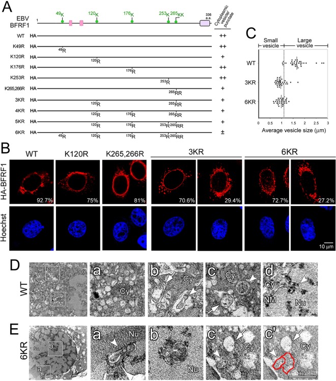
FIG 4.
Lys-to-Arg substitution of EBV BFRF1 influences its NE-modulating and vesicle-forming abilities. (A) Schematic representation of HA-tagged BFRF1 mutants with multiple Lys-to-Arg substitutions. Putative late domains (pink boxes) and a transmembrane domain (purple box) were predicted manually or by the MUSCLE and DAS programs. Six putative ubiquitination sites (green dots), Lys-49, -120, -176, -253, -265, and -266, on BFRF1 were identified by the UbPred program. The abilities of BFRF1 mutants to induce cytoplasmic vesicles, or puncta, and intranuclear puncta are summarized on the right. Pluses and minuses indicate the relative qualifications of specific phenotypes in each experiment. (B) Confocal images of HA-BFRF1 WT-, F1(120R)-, F1(K265,266R)-, F1(3KR)-, and F1(6KR)- or control vector-transfected HeLa cells immunostained for HA and emerin. The vesicle morphology and emerin distribution in HA-BFRF1 WT- and mutant-expressing cells were observed. The percentages of cells with specific BFRF1 vesicle expression patterns were obtained from three independent experiments with more than 100 cells containing HA-BFRF1. (C) The average diameters of cytoplasmic vesicles in BFRF1 WT-, 3KR-, and 6KR-transfected cells was calculated in randomly selected sections with more than 20 cells in each set. Thirty-five randomly selected vesicles/puncta are shown with the average sizes indicated. The cutoff value for the average vesicle size was defined as 1.1 μm. (D and E) NE and vesicle ultrastructure in HA-BFRF1 WT- and F1(6KR)-expressing HeLa cells. (D) Expression of HA-BFRF1 WT altered the nuclear membrane (NM) structure and induced multiple irregular vesicles in the cytoplasm. The accumulation of cytoplasmic vesicles (a) or invagination of the NM (d, arrowhead) was observed in the HA-BFRF1 WT-transfected cells. Empty vesicles with single- or double-layer membranes (b and c, arrows) or vesicles containing fuzzy inclusions (b and c, arrowheads) were also found at higher magnification in cells transfected with HA-BFRF1. (E) Expression of F1(6KR) induced protrusion of the NM into the nucleus and multiple vesicles in the cytoplasm. The modulated NM (a, arrowhead), intranuclear vesicle with double membranes (b), and cytoplasmic vesicle without (c and c′, arrows) or with (c and c′, arrowheads and red line) fuzzy inclusions are shown at higher magnification. The boxed regions in the left image are enlarged on the right.

