FIG 7.
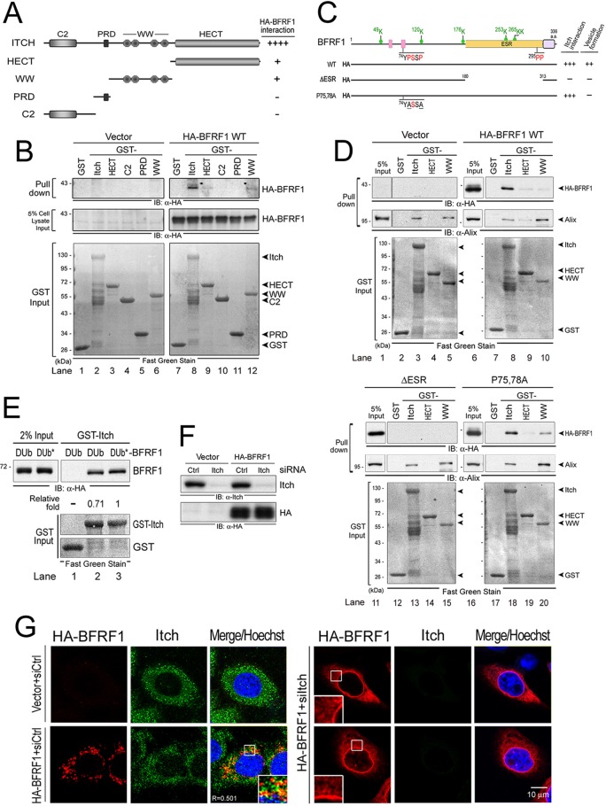
The ESR of BFRF1 mediates Itch complex association. (A) Schematic representation of GST-fused Itch fragments. The relative association intensities between the GST-fused Itch fragments and HA-BFRF1 are summarized on the right. Pluses and minuses indicate the relative qualifications of specific phenotypes in each experiment. (B) Characterization of the BFRF1-interacting domain on Itch in vitro. Equal amounts of bacterially expressed GST, GST-fused full-length Itch, HECT, WW, PRD, and the C2 domain fragment were captured by glutathione beads and incubated with the HA-BFRF1-expressing cell lysates. Inputs of HA-BFRF1 and GST-fused proteins were detected by HA antibody and Fast Green staining, respectively. (C) Schematic representation of HA-tagged BFRF1 mutants for mapping the Itch-interacting region. Putative late domains (pink boxes), the transmembrane domain (purple box), and putative ubiquitination sites (green dot) are predicted. The Itch-interacting abilities of BFRF1 mutants in GST pulldown analysis are indicated on the right. Red letters in the sequences indicate the putative Itch-targeting residues; underlined letters indicate mutated residues. (D) Bacterially expressed GST-Itch, GST-HECT, GST-WW, and GST fragment were captured by glutathione beads and incubated individually with the lysates from HA-BFRF1 WT-, F1(ΔESR)-, F1(P75,78A)-, or vector plasmid-transfected HeLa cells. The pulled down protein complexes were then detected by antibodies against HA and Alix. The GST protein inputs were detected by Fast Green staining. (E) Bacterially expressed GST-Itch and the GST fragment were captured by glutathione beads and incubated individually with the lysates from DUb- or Dub*-BFRF1 plasmid-transfected HeLa cells. The pulled down protein complexes were then detected by antibodies against HA. The GST protein inputs were detected by Fast Green staining. (F) Lysates from HeLa cells transfected with Itch or control (Ctrl) siRNA, together with a plasmid expressing HA-BFRF1 or a control vector, were harvested at 72 h posttransfection and subjected to immunoblot analysis using antibodies against Itch and HA. (G) HeLa cells were transfected with Itch (siItch) or control (siCtrl) siRNA for 48 h and cotransfected with siRNAs and a plasmid expressing HA-BFRF1 for an additional 24 h. HA (red) and Itch (green) were visualized by indirect immunofluorescence and observed by confocal microscopy. The boxed regions are enlarged in the insets.

