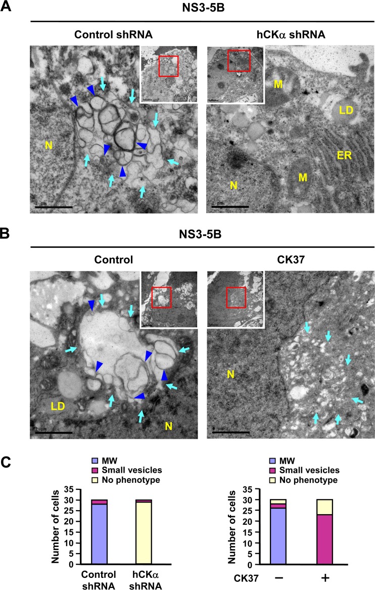
FIG 7.
Abrogation of HCV-induced MW formation by hCKα depletion or CK37 treatment. (A) Control shRNA and hCKα shRNA stable expression cells were transfected with pTM-NS3-5B and pWPI-T7-BLR, fixed, and processed for TEM. (B) Huh7 cells were first cotransfected with plasmids expressing T7 polymerase and NS3-NS5B. At 24 h post-DNA transfection, cells were left untreated or treated with 100 μM CK37 for 18 h, and then cells were analyzed by TEM for MW formation. The area boxed in red was enlarged as shown. Light blue arrows indicate vesicles of heterogeneous sizes (left panels of A and B) and small vesicles in CK37-treated Huh7 cells (right panel of B); blue arrowheads indicate double-membrane vesicles (DMVs) (left panels of A and B). N, nucleus; LD, lipid droplet; ER, endoplasmic reticulum; M, mitochondria. (C) Thirty cells randomly picked from each experimental setting were examined by TEM, and the numbers of cells showing three different phenotypes were quantified. MW, vesicles 130 to 270 nm in diameter; small vesicles, vesicles 40 to 100 nm in diameter.

