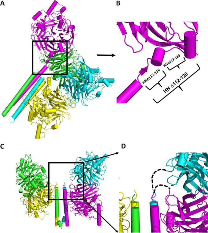
FIG 1.

HN structure with head-stalk connecting loops. (A) Structure of PIV5 virus HN showing “two heads up” and “two heads down” with four helix bundle stalk conformation (PDB 4JF7) (61) shown in a 45° slanted representation. Each protomer is colored differently. (B) Expanded section of a single protomer in panel A showing connecting “flexible” helix and loops. The deletion mutations made are indicated by brackets. (C) Structure of NDV HN (PDB 3T1E) showing four heads down arrangement of the head and stalk. (D) Expanded section of panel C showing missing residues connecting heads to stalks indicated by dashed lines.
