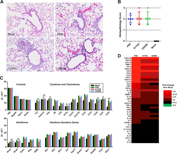
FIG 7.
Host responses to A- and B-allele segment 8 reassortant viruses. (A) At day 6 p.i., the right lung lobes of inoculated mice were collected, fixed, processed, and stained with hematoxylin and eosin. Mock-infected mice showed no significant changes, whereas infected mice showed degeneration and necrosis of epithelial cells lining the airways with peribronchiolar and perivascular inflammation and interstitial inflammation sometimes accompanied by necrosis and fibrin accumulation. The inflammatory infiltrate was predominately lymphocytes and macrophages with variable numbers of neutrophils. Bars, 50 μm. (B) The severity of the pathology in the lung was assessed in a blind manner, and an overall pathology score out of 3 was assigned. (C) RNA was extracted from the lungs of infected mice at day 4 p.i., and the levels of various cytokines, chemokines, and antiviral gene expression were quantified using RT-qPCR. Data are plotted as the mean (20 − dCT) ± SD. The values for all infected samples were significantly different from the value for the mock-infected sample, unless the bar is labeled with NS, which indicates no significant difference by the unpaired t test. *, P < 0.05; **, P < 0.01. (D) The levels of various cytokines and chemokines in pooled lung homogenates from groups of 5 mice culled at day 4 p.i. were determined using an immunoblot spot array. Values are represented by those from a heat map of the fold change with respect to the level of expression for the mock-infected samples.

