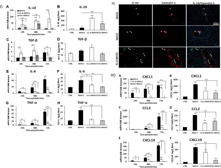
FIG 3.
Gene expression and production of IL-10, TGF-β, IL-6, TNF-α, CXCL1, CCL2, and CXCL10 in the livers of MHV3-, 51.6-MHV3-, and YAC-MHV3-infected mice. Groups of 5 or 6 C57BL/6 mice were intraperitoneally infected with 1,000 TCID50 of MHV3, 51.6-MHV3, and YAC-MHV3. At 24, 48, or 72 h p.i., livers were collected from mock- and virus-infected mice of each group. (I) Fold changes in mRNAs of IL-10 (A), TGF-β (C), IL-6 (E), and TNF-α (G) were analyzed by qRT-PCR. Values represent fold change in gene expression relative to those in mock-infected mice (arbitrarily taken as 1) after normalization to HPRT expression. Production levels of IL-10 (B), TGF-β (D), IL-6 (F), and TNF-α (H) at 72 h p.i. in the liver of each mouse were quantified by ELISA. (II) In situ expression of IL-10 and caveolin-1 was assayed by immunohistochemistry in livers of mock-, MHV3-, and 51.6-MHV3-infected mice at 48 h p.i. (arrows show IL-10- and caveolin-1-expressing endothelial cells). (III) Fold changes in mRNAs of CXCL1 (A), CCL2 (C), and CXCL10 (E) were analyzed by qRT-PCR. Values represent fold change in gene expression relative to that in mock-infected mice (arbitrarily taken as 1) after normalization to HPRT expression. Production levels of CXCL1 (B), CCL2 (D), and CXCL10 (F) at 72 h p.i. in the liver of each mouse were quantified by ELISA. Values are means and standard errors of the means. *, P < 0.05; **, P < 0.01; ***, P < 0.001 (compared with mock-infected mice). †, P < 0.05; ††, P < 0.01; †††, P < 0.001 (compared with MHV3-infected mice).

