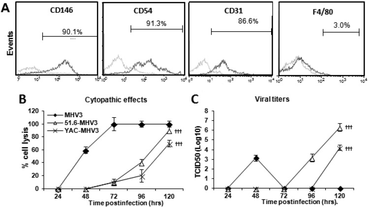
FIG 6.
Permissivity of LSECs to MHV3, 51.6-MHV3, and YAC-MHV3 infection. Mouse LSECs were isolated with Percoll gradients and enriched by immunomagnetism with anti-CD146 antibodies. (A) LSECs were characterized by immunolabeling with antibodies for CD146, CD54, CD31, and F4/80 cell markers and cytofluorometric analysis. (B and C) LSECs were infected at an MOI of 0.1 with MHV3, 51.6-MHV3, and YAC-MHV3. The evolution of cytopathic effects in LSEC cultures was noted up to 5 days p.i. (B), and the kinetics of MHV infections were monitored by quantifying viral titers in supernatants of infected LSECs (C). All experiments were conducted in triplicate. Results are representative of two independent experiments. Values are means for each point. †††, P < 0.001 compared with MHV3-infected cells.

