
Figure 1.
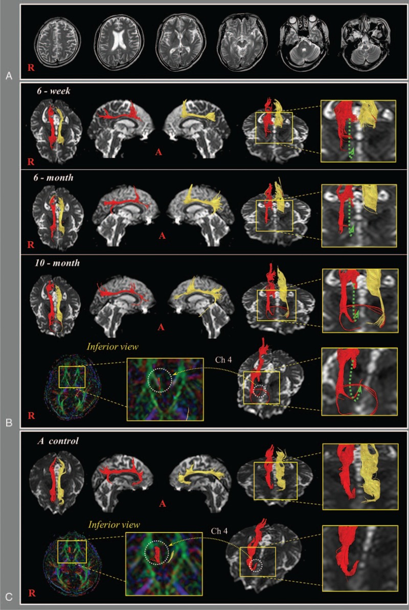
(A) Brain magnetic resonance imaging at 6 weeks after onset showed a subdural hygroma located in the left frontal convexity. (B) DTTs of the patient; 6-week DTT: discontinuation (green arrows) superior to the genu of the corpus callosum is observed in both cingulums, 6-month DTT: the discontinued anterior part of the right cingulum is elongated anteriorly through the anterolateral subcortical white matter of the cingulum (blue arrow), 10-month DTT: this elongated neural tract of the right cingulum is connected to the right nucleus basalis of Meynert (Ch 4) in the basal forebrain. (C) DTT images of the cingulum in a normal control subject (48-year-old male). DTT = diffusion tensor tractography.