Figure 2.

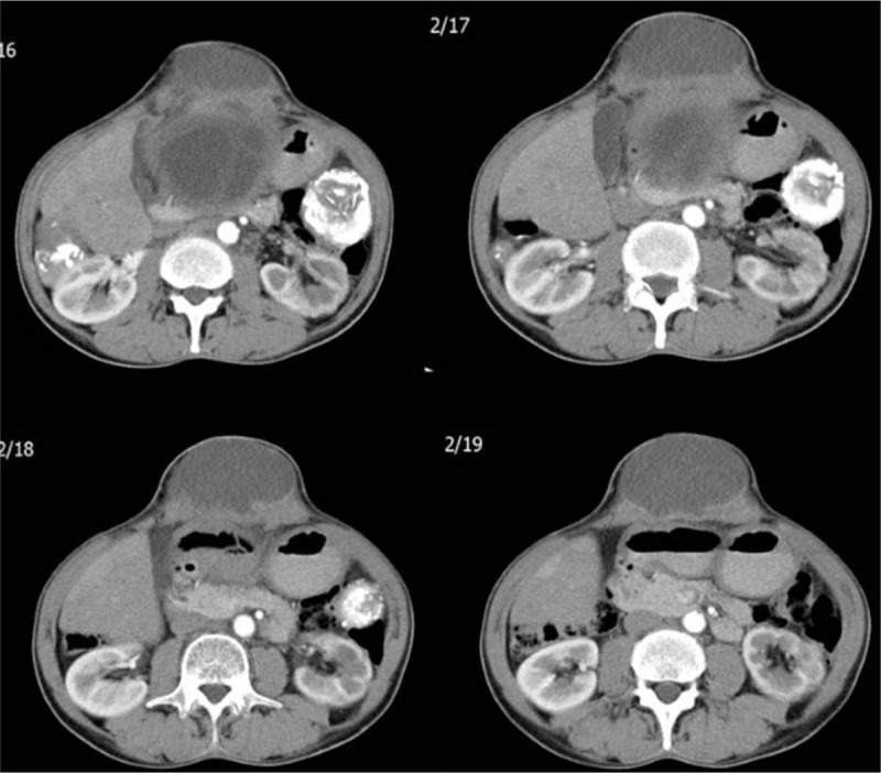
Sequential abdominal computed tomography images taken after injecting contrast material. A lesion consistent with a hydatid cyst is seen starting from segment 3 of the left liver lobe and protruding into the anterior abdominal wall.

Sequential abdominal computed tomography images taken after injecting contrast material. A lesion consistent with a hydatid cyst is seen starting from segment 3 of the left liver lobe and protruding into the anterior abdominal wall.