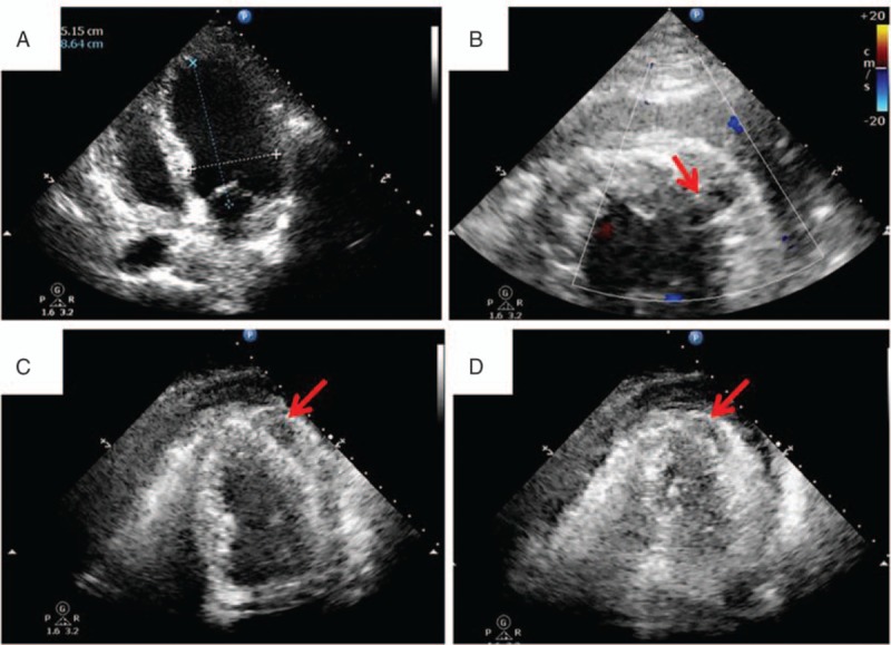
Figure 2.

Preoperative cardiac images, (A) transthoracic echocardiography before the radiofrequency catheter ablation (RFCA), (B) transthoracic echocardiography after the RFCA (red arrow: rupture of the left ventricular wall), (C) transthoracic echocardiography after the RFCA (red arrow: hematoma in the left ventricular wall during diastole), (D) transthoracic echocardiography after the RFCA (red arrow: hematoma in the left ventricular wall during systole: smaller than that during diastole due to the contraction of the myocardium).
