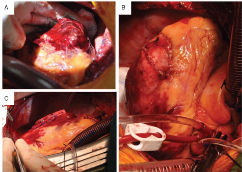
Figure 3.

Intraoperative cardiac images, (A) a 6 × 8 cm2 area of contusion in the posterolateral wall was found. At the center of the contusion near the apex, a 5-cm tear of the epicardium was revealed, and 1 cm of the tear penetrated into the whole wall. (B) The rupture of the left ventricular wall was repaired by teflon-buttressed sutures. (C) A pericardium patch of sufficient size was applied to overlay the contusion region with interrupted sutures around and bioglue inside.
