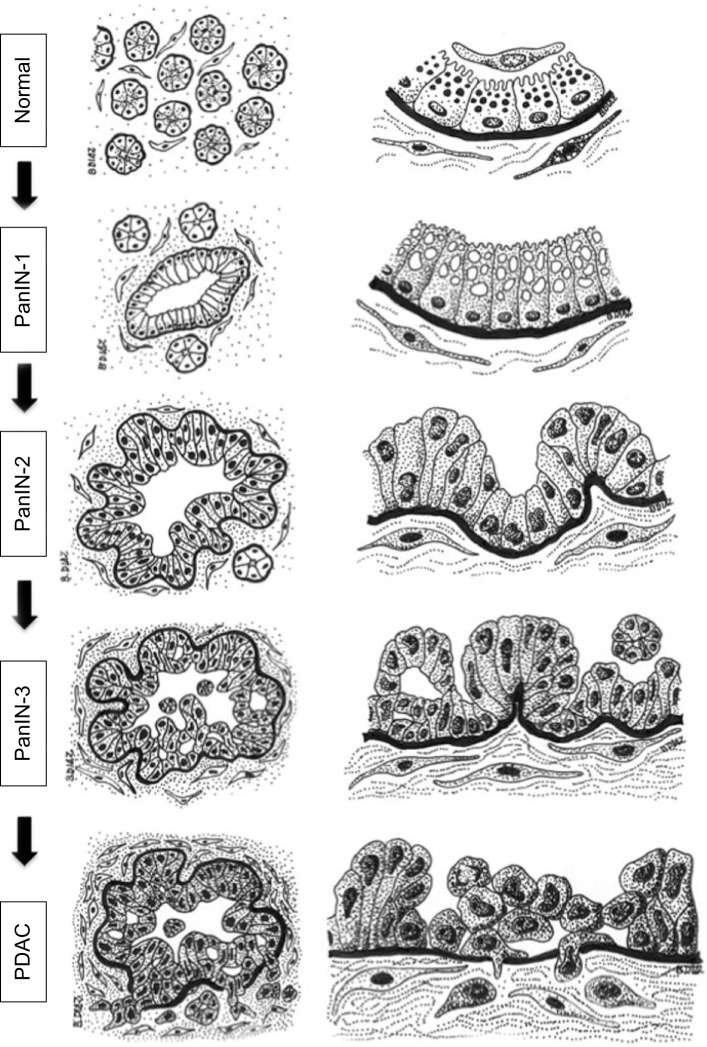
Figure 2.
Diagram of the histology of precursor lesions and PDAC.
Notes: The progression model from normal exocrine pancreas to PDAC is depicted as diagrams of representative tissue sections (left column) and their corresponding magnifications (right column). The normal exocrine pancreas is formed by pancreatic acini surrounded by stromal tissue containing fibroblasts and pancreatic stellate cells. Each acinus contains ductal (not depicted), centroacinar, and acinar cells that contain zymogen granules and are separated from the stroma by a basement membrane (black). Recent studies indicate that acinar cells may be the epithelial cell type targeted by oncogenic transformation during PDAC initiation, although a centroacinar or ductal origin for PDAC cannot be completely ruled out (see text for details). PanIN lesions already contain mutations in K-RAS and are characterized by a columnar epithelium resembling the ductal epithelium. Cells in PanIN-1 often contain mucin in the cytoplasm and present normal nuclei located basally. PanIN-2 lesions are characterized by hyperplasia (excessive cell proliferation), which leads to papillary protrusions into the lumen. The epithelium often becomes pseudostratified, and not all nuclei are basally located, indicating initial loss of epithelial polarity. Nuclei are abnormally shaped. PanIN-3 is the most advanced precursor lesion and displays extensive hyperplasia with papillary protrusions into the lumen, loss of epithelial polarity, and abnormally shaped nuclei that pile up. In PDAC, cells become mesenchymal and invasive, cross the basement membrane, and migrate through the stroma. The integrity of the basement membrane is the main histological feature that differentiates PanIN-3 and PDAC, although recent studies indicate that cells may cross the basement membrane earlier during the progression of the disease (see text for details). Note that the stroma surrounding the lesions and the cancer cells also evolves during the progression of PDAC, becoming more dense and fibrotic. Fibrosis starts in PanIN-1 lesions and progressively increases through PanIN-2 and -3 until the formation of the intense desmoplastic reaction that characterizes PDAC (see text for details).
Abbreviations: PanIN, pancreatic intraepithelial neoplasia; PDAC, pancreatic ductal adenocarcinoma.

