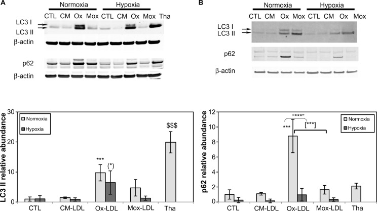
Figure 5.
Effects of hypoxia on low-density lipoprotein-induced autophagy.
Notes: Macrophages were incubated for 72 hours with 200 μg/mL of oxidized/modified low-density lipoproteins under hypoxia (1% oxygen) or normoxia (21% oxygen) or with thapsigargin at 200 nM for 20 hours under normoxia for (A) THP-1-derived macrophages or (B) peripheral blood monocytic cell-derived macrophages. The abundance of LC3 and p62 was assessed by Western blot analysis from total protein extracts using specific antibodies. In both cases, β-actin was used as the loading control. Histograms show, for THP-1-derived macrophages, the ratio of the intensity of the LC3 II or p62 band related to the intensity of the β-actin band as the mean ratio of three independent experiments ± standard deviation. ***P<0.001 versus normoxic control. (*)P<0.05 versus hypoxic control. “***”P<0.001 versus corresponding normoxic cells. [***]P<0.001 myeloperoxidase-modified versus copper sulfate-oxidized low-density lipoproteins. $$$P<0.001 thapsigargin versus control using Student’s t-test.
Abbreviations: CM(-LDL), cell-modified/native (low-density lipoproteins); CTL, control; H, hypoxia; Mox(-LDL), myeloperoxidase-modified (low-density lipoproteins); N, normoxia; Ox(-LDL), copper sulfate-oxidized (low-density lipoproteins); Tha, thapsigargin.

