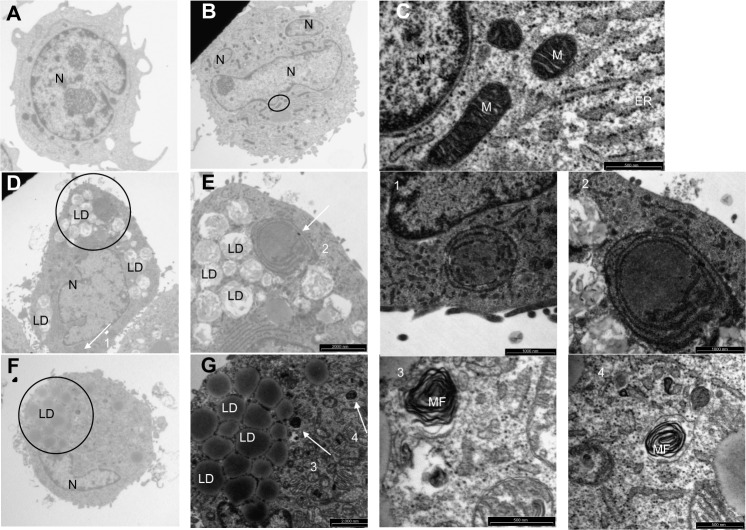
Figure 6.
Effects of low-density lipoproteins on cell morphology.
Notes: Macrophages were incubated for 72 hours with or without 200 μg/mL of low-density lipoproteins under normoxia (21% oxygen). After incubation, the cells were fixed and processed for analysis by transmission electron microscopy. (A and B) Cells incubated for 72 hours in normoxia without low-density lipoproteins (magnification 2,550×). (C) Enlargement of the area circled in (B) (magnification 30,000×). (D) Cells incubated for 72 hours under normoxia with copper sulfate-oxidized low-density lipoproteins (magnification 2,550×). (E) Enlargement of the area circled in (D) (magnification 6,200×). (F) Cells incubated under normoxia for 72 hours with myeloperoxidase-modified low-density lipoproteins (magnification 2,550×). (G) Enlargement of area circled in (F) (magnification 6,200×). (1) Enlargement of the structure pointed to by Arrow 1 in (D) (magnification 15,000×). (2) Enlargement of the structure pointed to by Arrow 2 in (E) (magnification 30,000×). (3) Enlargement of the structure pointed to by Arrow 3 in (G) (magnification 30,000×). (4) Enlargement of the structure pointed to by Arrow 4 in (G) (magnification 30,000×).
Abbreviations: ER, endoplasmic reticulum; LD, lipid droplets; M, mitochondria; MF, myelin figures; N, nucleus.

