Figure 2.

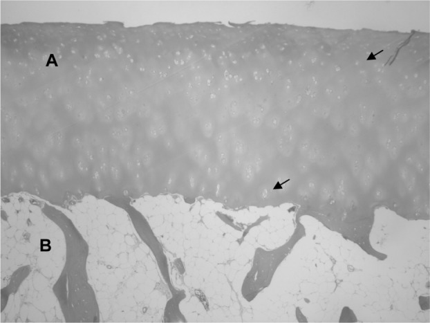
Normal histological appearance of the hyaline cartilage (A) and the subchondral bone (B).
Note: Arrows indicate chondrocytes inside their lagoons.

Normal histological appearance of the hyaline cartilage (A) and the subchondral bone (B).
Note: Arrows indicate chondrocytes inside their lagoons.