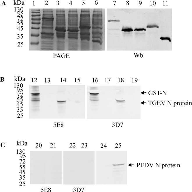
Fig 1. Preparation of mAbs against the N protein of TGEV.
(A) Expression and purification of TGEV GST-N, GST-N1, GST-N2 and GST-N3 proteins. The proteins were visualized using PhastGel Blue R staining (lanes 1–6) or were detected after western blotting with a GST mAb (lanes 7–11). Lanes 2 and 7: GST-N protein. Lane 1: protein molecular weight marker. Lanes 3 and 8: GST-N1 protein. Lanes 4 and 9: GST-N2 protein. Lanes 5 and 10: GST-N3 protein. Lanes 6 and 11: GST protein. (B) Reactivity of the mAb 5E8 with the GST-N protein and the TGEV N protein. Lanes 12 and 16: GST-N protein. Lanes 13 and 17: GST protein. Lanes 14 and 18: cell lysates of TGEV-infected PK-15 cells. Lanes 15 and 19: cell lysates of mock-infected PK-15 cells. (C) Reactivity of 5E8 and 3D7 with PEDV N protein. Lanes 20, 22, and 24: cell lysates of PEDV-infected Vero E6 cells. Lanes 21, 23, and 25: cell lysates of mock-infected Vero E6 cells. The PEDV mAb was maintained in the lab.

