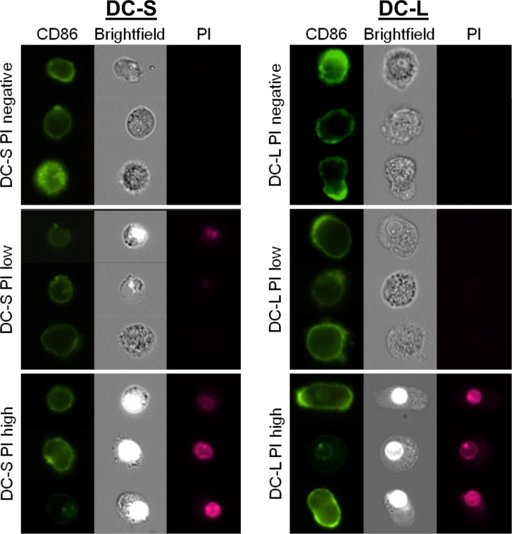
Fig 6. Imaging of live DCs stained with CD86 and PI.
iDCs were labeled with CD86, co-stained with PI and imaged using an Amnis Imagestream™ cytometer. The cells were gated into DC-S (left column) and DC-L (right column), as well as PI-negative, low, and high, using an analogous scheme to the one used with other flow cytometers. Three representative examples from every set are shown.

