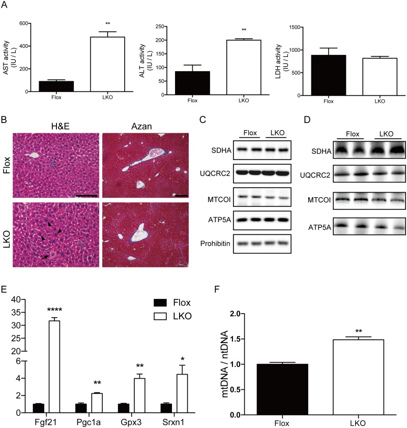
Fig 8. Sustained liver function in adult Mtu1-deficient mice.
(A) Serum AST, ALT and LDH levels in LKO and Flox mice at the age of 16 weeks. n = 3 per genotype, **P < 0.01. (B) H&E and Azan staining in 16-week-old LKO and Flox mice. The arrow indicates spotty necrosis. Arrowheads indicate enlarged hepatocytes with karyomegaly. Bar = 0.2 mm for H&E staining. Bar = 0.1 mm for Azan staining. (C) Protein levels of representative mitochondrial proteins in the livers of 16-week-old Flox and LKO mice were examined by western blotting. (D) Mitochondrial proteins incorporated in Complexes I ~ V were examined by blue-native PAGE followed by western blotting. (E) The relative expression levels of Fgf21, Pgc1a, Gpx3 and Srxn1 in the livers of Flox and LKO mice are shown. n = 3 each. *P < 0.05, **P < 0.01, ****P < 0.0001. (F) The ratios of mtDNA levels to ntDNA levels in the livers of 16-week-old Flox and LKO mice are shown. n = 3 each. **P < 0.01.

