. 2016 Sep 8;5:e20125. doi: 10.7554/eLife.20125

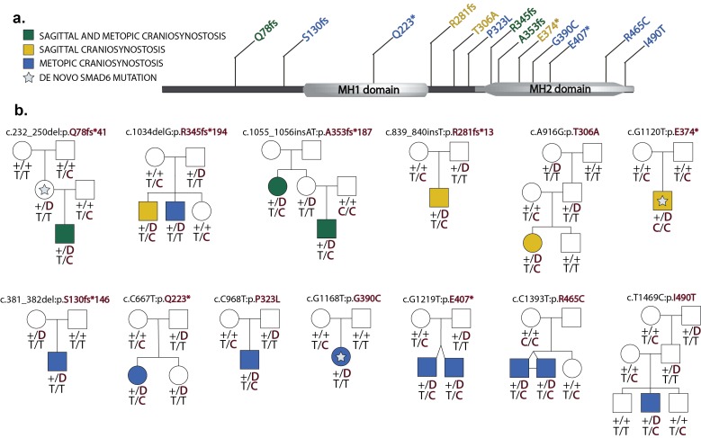
Figure 2. Segregation of SMAD6 mutations and BMP2 SNP genotypes in pedigrees with midline craniosynostosis.

(a) Domain structure of SMAD6 showing location of the MH1 and MH2 domains. The MH1 domain mediates DNA binding and negatively regulates the functions of the MH2 domain, while the MH2 domain is responsible for transactivation and mediates phosphorylation-triggered heteromeric assembly with receptor SMADs. De novo or rare damaging mutations identified in craniosynostosis probands are indicated. Color of text denotes suture(s) showing premature closure. (b) Pedigrees harboring de novo (denoted by stars within pedigree symbols) or rare transmitted variants in SMAD6. Filled and unfilled symbols denote individuals with and without craniosynostosis, respectively. The SMAD6 mutation identified in each kindred is noted above each pedigree. Below each symbol, genotypes are shown first for SMAD6 (with 'D' denoting the damaging allele) and for rs1884302 risk locus downstream of BMP2, (with 'T' conferring protection from and 'C' conferring increased risk of craniosynostosis). All 17 subjects with craniosynostosis have SMAD6 mutations, and 14/17 have also inherited the risk allele at rs1884302, whereas only 3 of 16 SMAD6 mutation carriers without the rs1884302 risk allele have craniosynostosis.

DOI: http://dx.doi.org/10.7554/eLife.20125.006

Figure 2—source data 1. Variants identified in SMAD6.
Highlighted variants indicate de novo mutations; 'D' and 'T' respectively denote damaging and tolerated missense variants called by MetaSVM.
DOI: 10.7554/eLife.20125.007
Figure 2—source data 2. PCR primer sequences for Sanger sequencing of reported variants.
DOI: 10.7554/eLife.20125.008

Figure 2.

Figure 2—figure supplement 1. Plots of independent Illumina sequencing reads in a parent-offspring trio showing de novo SMAD6 mutation.
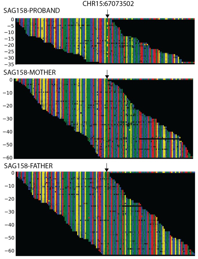
Figure 2—figure supplement 1.

The reference sequence of a segment of SMAD6 that includes base 15:67073502 (denoted by arrow) is shown in the top row, with red, blue, green and yellow squares representing A, C, G, T, respectively. Below, all independent reads that map to this interval are shown. The results show that the proband has 23 reads of reference ‘G’, and 10 reads of non-reference ‘T’. Only the reference ‘G’ is seen in both parents, providing evidence of a de novo mutation.
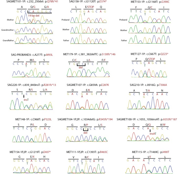
Figure 2—figure supplement 2. Confirmation of SMAD6 mutations by Sanger sequencing of PCR products.

Figure 2—figure supplement 2.

Sanger sequencing traces of PCR amplicons containing SMAD6 mutations identified by exome sequencing are shown. Above each trace or set of traces, the kindred ID, mutation identified in the DNA sequence and its impact on SMAD6 protein is indicated. Above sequence traces, the inferred DNA sequence is shown, along with the inferred amino acid sequence (shown in single letter code). Heterozygous mutations are indicated beneath the wild-type sequence and non-reference amino acid sequences are shown in red. Deleted and inserted bases are denoted, and result in an overlap of wild-type and mutant sequences.