. 2016 Sep 8;5:e20125. doi: 10.7554/eLife.20125

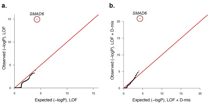
Figure 3. Quantile-quantile plots of observed versus expected p-values comparing the burden of rare LOF and damaging (LOF + D-mis) variants in protein-coding genes in craniosynostosis cases.

Rare (allele frequency <2 × 10⁻⁵ in the ExAC03 database) loss of function (LOF) and damaging missense (D-mis) variants were identified in 191 probands. The probability of the observed number of variants in each gene occurring by chance was calculated from the total number of observed variants and the length of the coding region of each gene using the binomial test. The distribution of observed P-values compared to the expected distribution is shown. (a) Q-Q plot for rare LOF variants in each gene from a total of 1135 LOF variants identified in probands. The distribution of observed p-values closely conforms to expectation with the exception of SMAD6, which shows p=1.1 × 10⁻¹⁵ and 156-fold enrichment in cases. (b) Q-Q plot for rare damaging (LOF + D-mis) variants in each gene from a total of 3156 damaging variants in probands. Again, SMAD6 deviates greatly from the expected distribution, with p<10–20 and 91-fold enrichment.

DOI: http://dx.doi.org/10.7554/eLife.20125.012

Figure 3—source data 1. Source data for Figure 3—figure supplement 3.
elife-20125-fig3-data1.docx (131.8KB, docx)
DOI: 10.7554/eLife.20125.013

Figure 3.

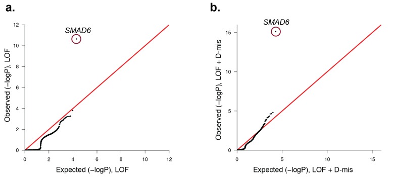
Figure 3—figure supplement 1. Quantile-quantile plots comparing all transmitted, damaging variants in protein-coding genes in 191 probands with midline craniosynostosis to the expected binomial distribution.

Figure 3—figure supplement 1.

De novo variants were excluded from this analysis, leaving 1122 rare (ExAC allele frequency < 2 x10⁻⁵), transmitted LOF variants and 3115 transmitted damaging (LOF + D-mis) variants. All genes closely matched expectation, with the exception of SMAD6. (a) There were 6 transmitted SMAD6 LOF mutations, a 118-fold enrichment compared to the expected 0.05 (p=2.2 × 10⁻¹¹). (b) Similarly, there were 10 transmitted damaging SMAD6 variants, a 71-fold enrichment compared to the expected 0.14 (p=7.0 × 10⁻¹⁶). The results demonstrate genome-wide significance of rare transmitted variants in SMAD6 independent of de novo mutations.
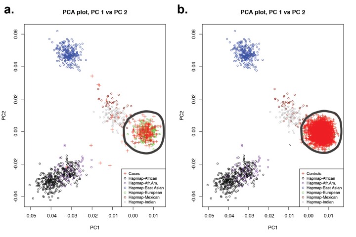
Figure 3—figure supplement 2. Principal-component analysis of 191 probands and 3337 European autism controls.

Figure 3—figure supplement 2.

(a) Principal component analysis of exome sequence genotypes from 191 probands with sagittal, metopic, or combined sagittal and metopic craniosynostosis clustered along with HapMap subjects. Results identify 172 craniosynostosis subjects that cluster with HapMap European subjects. (b) Principal component analysis of genotypes from exome sequencing data of European autism parent controls (n = 3337) showing clustering with HapMap subjects. In both panels, subjects considered to be of European ancestry are circled.
Figure 3—figure supplement 3. Quantile-quantile plot of observed versus expected p-values comparing the burden of damaging (LOF + D-mis) variants in protein-coding genes in craniosynostosis cases and controls.
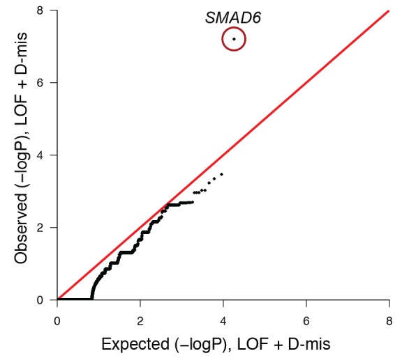
Figure 3—figure supplement 3.

The frequency of rare (allele frequency < 2 × 10⁻⁵ in the ExAC03 database) loss of function and D-mis variants in each gene was compared in 172 European probands with midline craniosynostosis and 3337 European controls. The distribution of observed p-values conforms to expectation with the exception of SMAD6, which deviates significantly from expectation. Because exon 1 of SMAD6 was poorly captured using the V2 capture reagent (used in control samples), 3 damaging variants in exon 1 in cases were excluded from this analysis.