. 2016 Sep 7;5:e15477. doi: 10.7554/eLife.15477

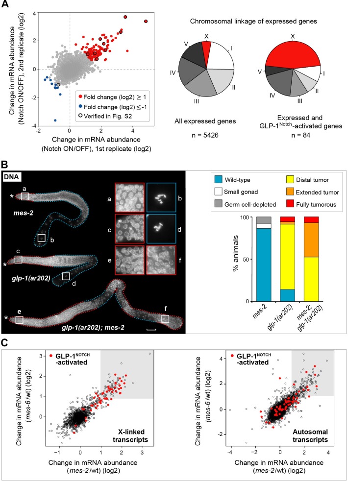
Figure 2. GLP-1Notch and PRC2 regulate common targets and are functionally connected.

(A) Notch-activated genes are biased for the sex chromosome linkage. Left: Changes in transcript abundance in the ‘Notch ON’ versus ‘OFF’ dissected gonads (genotypes explained in Figure 2—figure supplement 1A–B) were analyzed by microarrays. Transcripts upregulated at least 2-fold in the ‘Notch ON’ gonads are marked in red, those downregulated at least 2-fold in blue. Selected transcripts verified by RT-qPCR in Figure 2—figure supplement 2A are additionally circled in black. Right: 5426 genes can be considered expressed in the gonad, based on the bimodal distribution of expression values. Only 3% of those expressed genes are X-linked. In contrast, nearly half (46%) of the expressed and Notch-activated transcripts are X-linked (see Figure 2—figure supplement 2B for numbers). (B) GLP-1Notch and PRC2 interact genetically. Left: DAPI-stained gonads from animals of the indicated genotypes. The mes-2(bn11) M+Z- single mutant gonads have wild-type appearance at 20°C. The glp-1(ar202) gain-of-function mutants have an almost wild-type appearance at this temperature, except for an extended proliferative zone in the gonad, referred to as 'distal tumor'. At the same temperature, mes-2(bn11) M+Z-; glp-1(ar202) double mutants developed germline tumors in 32/32 of the examined gonads. The insets show close-ups from the indicated gonadal regions: the distal-most regions contain undifferentiated, proliferative germ cells in all mutants (a, c, e). However, while the single mutants contain oocytes with characteristically condensed chromosomes in the proximal gonads (b, d), the proximal gonads of the double mutants harbor proliferative germ cells (f). Scale bar = 30 μm. Right: quantification of the phenotypes. 'Distal tumor' indicates the presence of an elongated distal proliferative zone (approximately ½ of the distal gonad arm). 'Extended' tumor indicates an extended distal tumor, few oocytes, and frequently also a proximal tumor. 'Fully tumorous' indicates the absence of all differentiated cell types except for sperm produced during larval development. (C) GLP-1Notch and PRC2 target the same genes on the X chromosomes. The plots correlate changes in gene expression in M+Z- mes-2 mutants with changes in gene expression changes in M+Z- mes-6 mutants. Results are shown separately for X-linked (left) and autosomal (right) transcripts. Notch-activated genes (red in Figure 2A) are marked in red. Lightly shaded areas indicate transcripts that are at least 2-fold upregulated. The overlap between transcripts upregulated by GLP-1Notch and transcripts upregulated by the loss of CePRC2 is highly significant, particularly for the X-linked genes. The significance of the correlation was measured by hypergeometric distribution; X-linked Notch-activated vs. mes-2 derepressed: p=1.31e-31; X-linked Notch-activated vs. mes-6 de-repressed: p=7.41e-25; autosomal Notch-activated vs. mes-2 derepressed: p=1.47e-22; autosomal Notch-activated vs. mes-6 de-repressed: p=1.8e-12.

DOI: http://dx.doi.org/10.7554/eLife.15477.008

Figure 2—source data 1. Microarray results.
(A) Microarray results of differentially expressed genes in Notch ON/OFF gonads. Figure 2—figure supplement 3 source data: Quantification of germlines with wild-type morphology versus germlines with tumors upon germline-autonomous RNAi against genetic interactors of PRC2. More details can be found in the corresponding figure legends.
DOI: 10.7554/eLife.15477.009

Figure 2.

Figure 2—figure supplement 1. Examining transcriptional effects of GLP-1Notch signaling.

Figure 2—figure supplement 1.

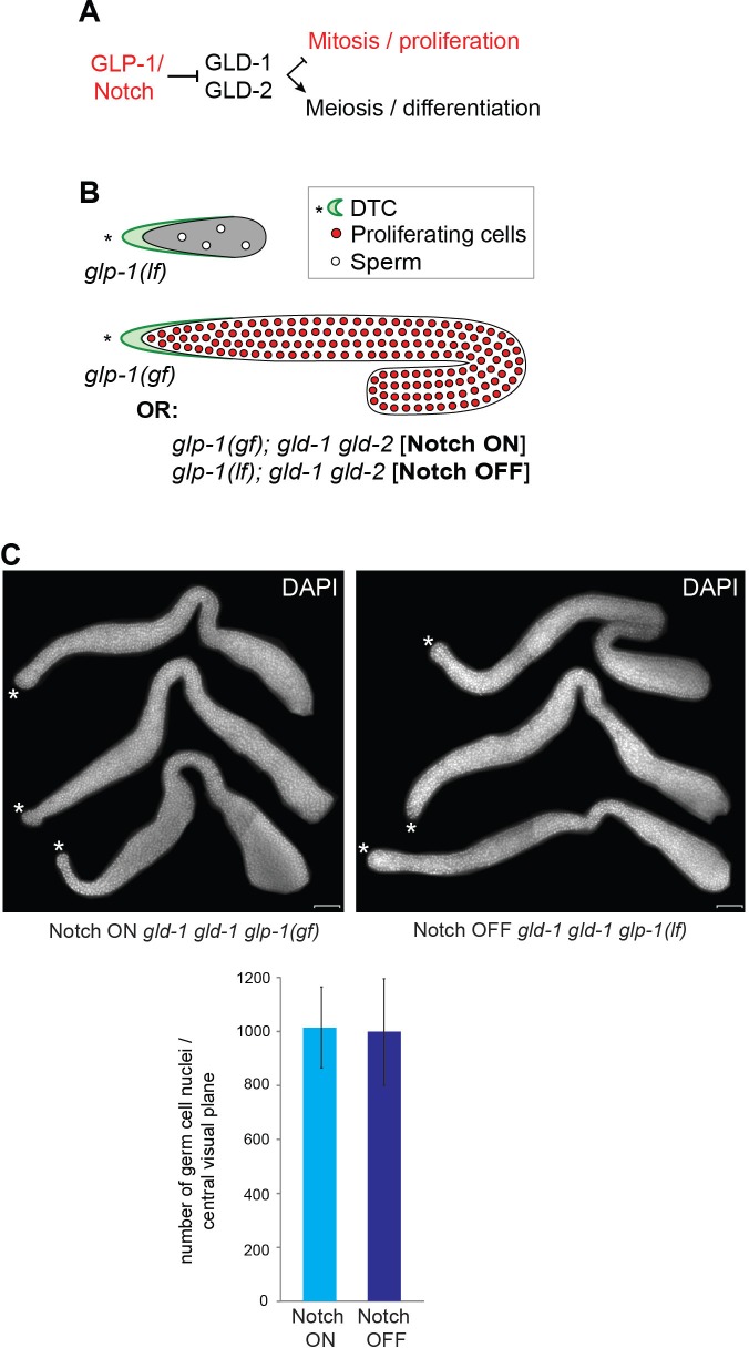
(A) GLP-1Notch signaling counteracts the meiosis-promoting activity of several RNA binding proteins, of which GLD-1 and -2 are indicated. (B) Mutants that were used in this study: while the loss-of-function (lf) alleles of glp-1 cause the loss of germ cells, gain-of-function (gf) alleles result in a constitutive proliferation of germ cells. However, in the absence of the meiosis-promoting GLD-1 and GLD-2 proteins, germ cells continue to proliferate in the absence of GLP-1Notch activity. (C) Three representative gonads of Notch ON: gld-2(q497) gld-1(q485); glp-1(ar202) and Notch OFF: gld-2(a497) gld-1(q485); glp-1(e2144) animals are shown after dissection and DAPI staining. The central planes of the gonads were imaged. Nuclei were counted from those images using the CellCounter plugin with ImageJ. For each genetic background, germ cells in the entire gonad of 10 dissected gonads were counted. The quantification below revealed that the numbers of germ cells in both backgrounds are not changed. Error bars represent SD. Scale bar = 10 μm.
Figure 2—figure supplement 2. Analysis of Notch-activated genes.

Figure 2—figure supplement 2.

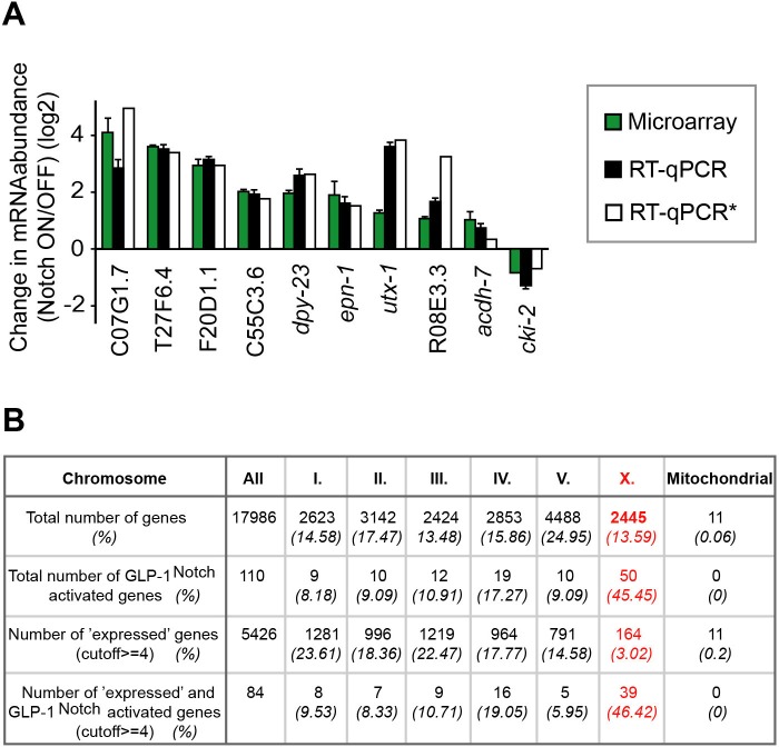
(A) Changes in the abundance of several transcripts (marked black in Figure 2A) were verified independently from the genomic quantification by RT-qPCR. Colors correspond to fold-changes detected by microarrays (green), by RT-qPCR on genetically identical gonads (black), or by RT-qPCR on gonads with a different loss-of-function glp-1 allele, q175 (white). *This experiment was performed only once. The error bars represent SEM. (B) Although the number of genes on the arrays is almost equally distributed between the different chromosomes, the expression of X-linked genes is largely silenced in the germline. The table displays the numbers and fractions of genes on the different chromosomes, and the distributions of the Notch-activated genes. The cutoff between 'not expressed' and 'expressed' genes was set according to the bimodal distribution of expression values in the Notch ON and OFF arrays.
Figure 2—figure supplement 3. The PRC2 component MES-6 and most enhancers/suppressors of glp-1(ar202) induced tumors appear to interact genetically with GLP-1Notch signaling in a germline-autonomous manner.

Figure 2—figure supplement 3.

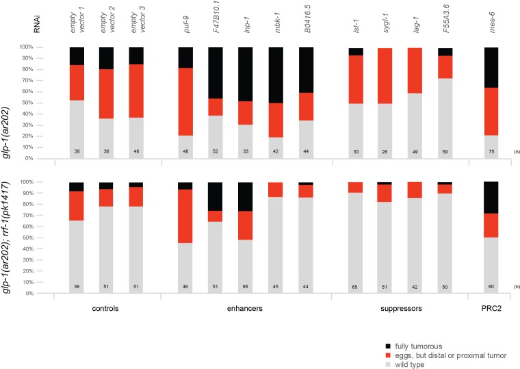
Each bar indicates the relative proportion of germlines with wild-type morphology (grey), germlines that contain eggs but also a proximal or distal tumor (red), and germlines that are fully tumorous (black). 'n' indicates the number of DAPI-stained gonads scored for each column. Empty vector 1–3 represent three independent replicates of the empty vector control and demonstrate the robustness of the experiment. We observed that, for an unknown reason, the rrf-1(pk1417); glp-1(ar202) double mutants were less likely to produce tumors at the semi-permissive temperature of 20°C. Nonetheless, the double mutant strain reacts to enhancers, suppressors, and depletion of PRC2 components in a similar manner as the glp-1(ar202) single mutant strain, with the exception of mbk-1 and B0416.5 RNAi. Depletion of mes-2 and mes-3 by RNAi was ineffective, since it did not enhance the tumourous phenotype in either of the two strains.
Figure 2—figure supplement 4. Global levels of H3K27me3 are unaffected by neither loss-of-function nor gain-of-function mutations in glp-1.

Figure 2—figure supplement 4.

Stainings of wild-type, gain-of-function glp-1(ar202), Notch ON (gld-2(q497) gld-1(q485); glp-1(ar202)) and Notch OFF (gld-2(a497) gld-1(q485); glp-1(q175)) gonads with antibodies against H3K27me3 performed on whole worms (left panel, scale bars = 10 μm) or dissected gonads (right panel, scale bars = 1 μm). The H3K27me3 levels do not differ globally between the different mutant backgrounds. Asterisks indicate distal tips of gonads, dashed lines outline gonad.