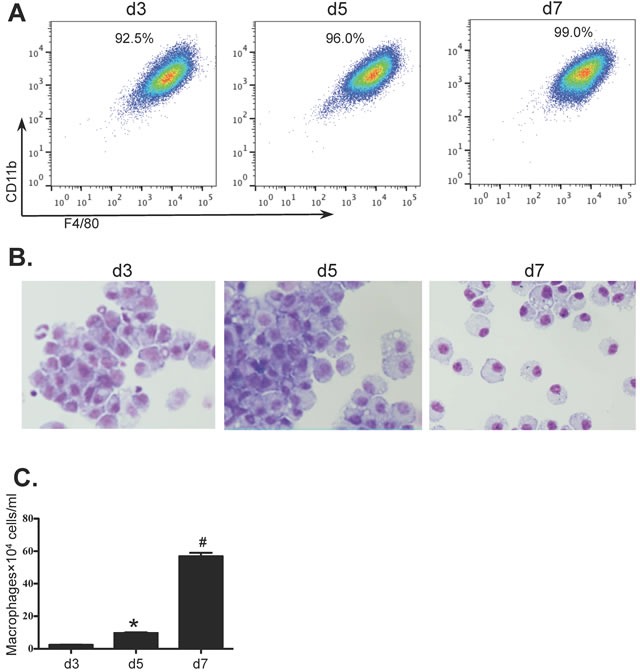
Figure 1. In-vitro differentiation of BMDM.

Bone marrow cells from BALB/c mice were cultured for 7 days (see Methods) and samples were collected on days 3, 5 and 7 from cultures grown in the presence of MCM. Macrophages were identified by A. flow cytometry (F4/80+CD11b+CD11c−Gr-1−), B. light microscopy with Giemsa staining (100×) and C. the numbers of BMDM were determined using a haemocytometer. Values are presented as mean ±SEM (n = 4~6), * P < 0.05 (v.s d3). # P < 0.05 (vs other groups).
