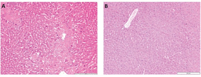
Figure 1.

A. H&E staining of Dkk2−/− liver tissue. Dkk2 deleted animals showed moderate to focally stronger variation of hepatocyte size with special regard to variable nuclear size and chromatin density. B. Corresponding wild type liver tissue with regular trabecular architecture and uniform hepatocytes.
