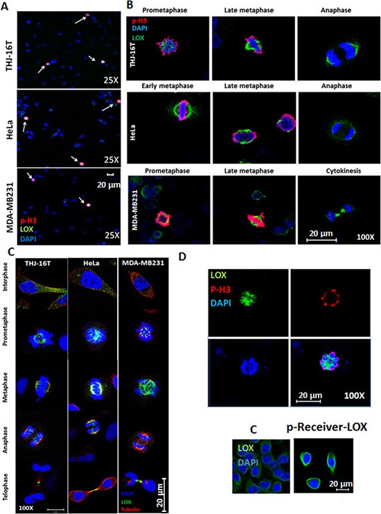
Figure 1. LOX is highly expressed in mitotic cells.

(A) LOX is highly expressed in p-H3(Ser10)-positive THJ-16T, HeLa, and MDA-MB231 cells (magnification 25X). (B) Subcellular localization of LOX in mitotic cells from prometaphase to anaphase (magnification 100X). (C) Co-localization of LOX and alpha tubulin on the mitotic spindles from metaphase to telophase (magnification 100X). (D) Top panel: p-H3(Ser10) and LOX staining in LOX-HeLa cells (magnification 120X). Bottom panel: Representative images of HeLa cells transfected with p-Receiver-LOX vector compared to the control cells (magnification 25X). C: Control cells.
