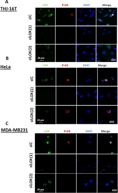
Figure 3. Knockdown of LOX protein using two independents siRNAs.

Immunofluorescence staining shows a decrease of LOX expression in siLOX (1) and siLOX (2) cells as compared to siControl in 3 cancer cells lines; THJ-16T (A), HeLa (B) and MDA-MB231 (C).
