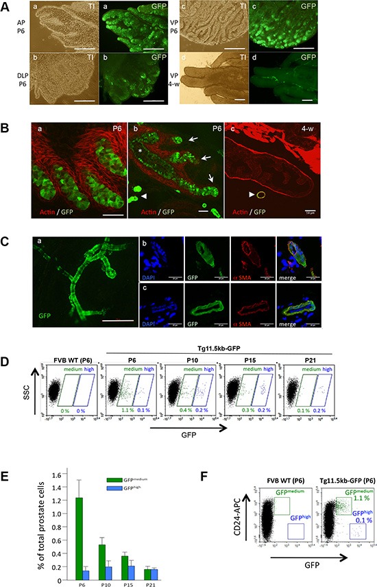
Figure 1. s-SHIP/GFP is transiently expressed in epithelial ducts during postnatal prostate development.

(A) Whole mount of anterior (AP), dorsolateral (DLP), and ventral (VP) prostate lobes of Tg 11.5kb-GFP mice, 6 days (P6) (a-c) or 4 weeks (4-w) (d) after birth were imaged under a fluorescence microscope. Representative picture (n = 3) of 4-w ventral lobe (d) is characteristic of all 4-w lobes. (B) Representative photographs (n > 10) of frozen sections of P6 (a,b) or 4-w (c) prostate tissues from Tg 11.5kb-GFP mice stained with phalloidin-Alexa594 for polymerized actin (red) to show the glandular architecture. (C) Representative photograph (n > 10) showing the typical morphology of blood vessels in Tg 11.5kb-GFP mice with GFP+-vascular smooth muscle cells (a). These vessel-associated GFP+ cells stained for alpha smooth muscle actin (b, c) (D) Representative flow cytometry analysis (n = 3) of GFP expressed by dissociated prostate cells isolated from P6 to P21 Tg 11.5kb-GFP mice. (E) Bar graph shows the frequency of GFPmedium (green bars) and GFPhigh (blue bars) cells in total dissociated prostate cells. Data represent the mean ± s.d., n = 3. (F) Representative flow cytometry (n > 5) analysis of GFP expressed by dissociated prostate cells isolated from P6 wild-type FVB (left panel) or P6 Tg 11.5kb-GFP (right panel) mice and labelled with anti-CD24 (APC) pan-epithelial marker. Transillumination (TI), side-scatter (SSC), allophycocyanin (APC), alpha smooth muscle actin (αSMA). Scale bars : 250 μm (A, Ca), 50 μm (B). 20 μm (Cb,c).
