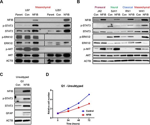
Figure 5. STAT3 signalling predicts NFIB function in GBM cell lines.

Changes in p-STAT3 but not ERK or AKT signalling paralleled the activity of NFIB in proneural, neural, classical and mesenchymal GBM cells - increased STAT3 phosphorylation was observed in (A) U87 and U251 (mesenchymal) GBM cells and in (B) classical and mesenchymal patient-derived GBM lines in response to increased NFIB expression. No change in p-STAT3 was observed in proneural cells and reduced expression was seen in neural GBM cells. In contrast no consistent correlation was observed between NFIB activity and either ERK or AKT signalling. (C) Expression of NFIB in the low-passage, unsubtyped GBM cell line Q1 was associated with increased expression of p-STAT3, increased expression of the astrocyte marker GFAP and (D) inhibition of cell proliferation.
