
Figure 6. CD109 knockdown in HUVEC facilitated tumor growth and metastasis in vivo.
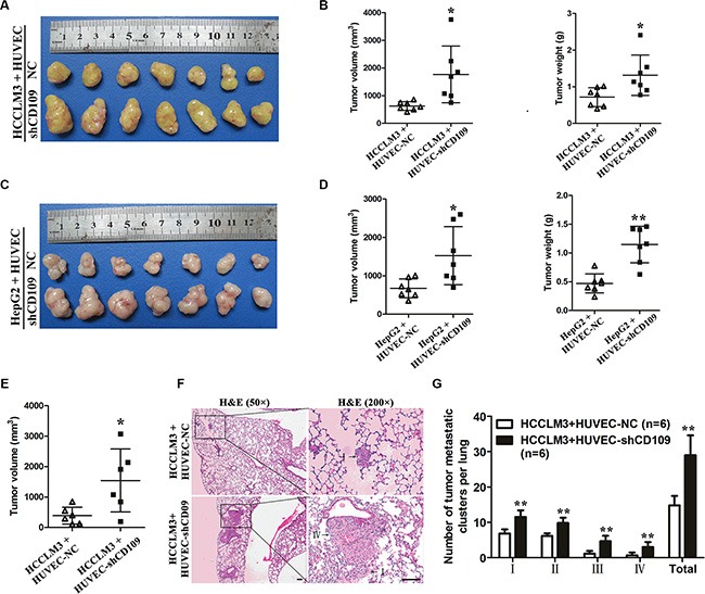
(A) Macrograph of subcutaneous tumors derived from HCCLM3 co-implantation with HUVEC-NC or HUVEC-shCD109. (B) Tumor volume and weight of HCCLM3-HUVEC co-implantation tumors were compared between the two groups (N = 7 mice per group). (C) Macrograph of subcutaneous tumors derived from HepG2 co-implantation with HUVEC-NC or HUVEC-shCD109. (D) Tumor volume and weight of HepG2-HUVEC co-implantation tumors were compared between the two groups (N = 7 mice per group). (E) Tumor volumes of HCCLM3-HUVEC co-implantation tumors were compared between the two groups in an orthotopic mouse model (N = 6 mice per group). (F) Representative images of lung metastatic foci with magnification of the selected areas (hematoxylin & eosin) were shown. Arrows indicated tumor grades. (G) The number and grade of lung metastatic foci were compared between the two groups (N = 6 mice per group). Scale bars, 100 μm. Data are presented as mean ± SD. *P < 0.05; **P < 0.01 by t test. NC, negative control.