
Figure 2. Generation of anti-CD3 × anti-B7-H3 bispecific antibody (B7-H3Bi-Ab).
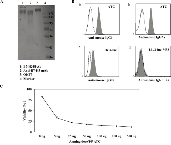
A. The heteroconjugated product of equimolar concentrations of B7-H3Bi-Ab was quantified by Coomassie blue staining of SDS-gel. B. Flow cytometry based binding assay for B7-H3Bi-Ab was tested. ATC was stained by B7-H3BiAb (shaded histogram), or a combination of OKT3 with anti-B7-H3 (the black line), then FITC-anti-mouse-IgG1 was added to detect the B7-H3 moiety of B7-H3Bi-Ab (a), or an FITC-anti-mouse IgG2a to detect the anti-CD3 moiety of the B7-H3Bi-Ab (b). (c) Hela-luc cells were incubated with B7-H3Bi-Ab (shaded histogram) or a combination of OKT3 and anti-B7-H3 (the black line), then B7-H3Bi-Ab binding was confirmed by FITC-anti-mouse IgG2a to detect the anti-CD3 moiety of the B7-H3Bi-Ab. (d) LL/2-luc-M38 cells were incubated with B7-H3Bi-Ab (shaded histogram) or a combination of OKT3 and anti-B7-H3 (the black line), then B7-H3Bi-Ab binding was analyzed by combined FITC-anti-mouse IgG2a with an anti-mouse-IgG1-FITC. C. Titer of B7-H3Bi-Ab armed ATC was measured. ATC was armed with B7-H3Bi-Ab ranging from 5 to 500 ng/106 cells at E/T ratio of 10:1, and cytotoxic effects of B7-H3Bi-armed ATC on Hela-luc cells were tested in vitro. After 18 hour incubation with B7-H3Bi-armed ATC, the percentage of viability was calculated at each concentration.