Fig. 2.
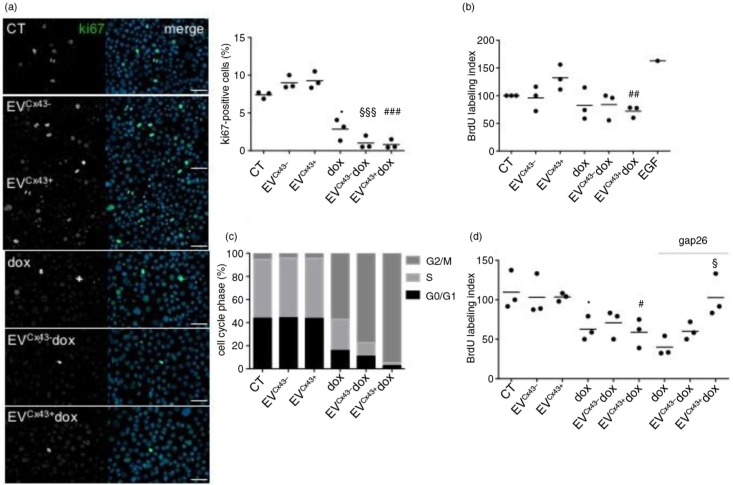
EVs loaded with dox impair tumour cell proliferation in vitro. 4T1luc2 cells were treated with 2 µM free dox, EVCx43+ dox, EVCx43− dox or vehicles, for 24 h. (a) Representative images of ki-67 immunostaining (green). Nuclei were stained with 4,6-diamidino-2-phenylindole (Dapi). Scale bars, 50 µm. Percentage of ki-67 (per total nuclei) is plotted in the graph (n=3, *p<0.05 vs. CT, §§§p<0.001 vs. EVCx43−, ###p<0.001 vs. EVCx43+). (b) Cell proliferation was assessed by the BrdU assay. Treatment with epidermal growth factor (100 ng/ml, 24 h) was performed as positive control. Percentage of BrdU incorporation is plotted on the graph (n=3, ##p<0.01 vs. EVCx43+). (c) Cell cycle analysis was performed by flow cytometry, using PI (n=3). (d) 4T1luc2 cells were treated with 2 µM free dox, EVCx43+ dox, EVCx43− dox or vehicles, for 1 h. A Cx43-mimetic peptide (gap26, 0.25 mg/ml) was used, where indicated. Fresh media was added and the cells cultured for an additional 23 h. Cell proliferation was assessed by the BrdU incorporation assay (n=3, *p<0.05 vs. CT, #p<0.05 vs. EVCx43+, §p<0.05 vs. EVCx43+ dox).

