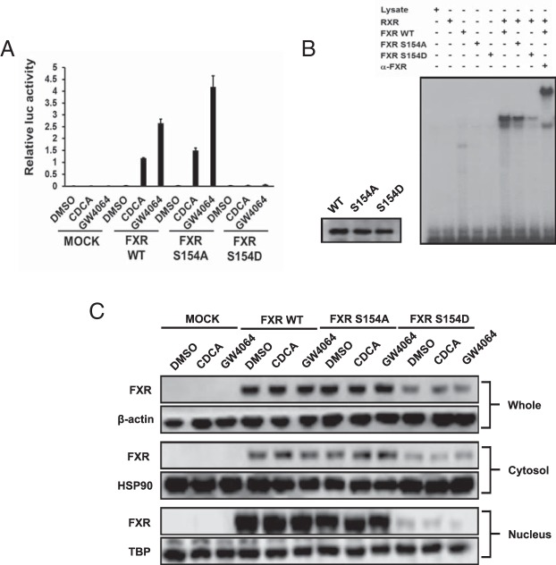
Figure 6. Functional characterization of hFXR mutants.
A, Cell-based reporter assay. Expression plasmids were cotransfected with a luciferase reporter construct for 24 hours. COS-1 cells were treated with 50μM CDCA and 5μM GW4064 for 24 hours. Empty expression plasmid pcDNA3.1 was used for mock transfection. Results are presented as mean ± SD of triplicate experiments. B, Gel-shift assays. The in vitro transcribed/translated proteins of the indicated hFXRs was incubated with 32P-labeled probe, electrophoresed on a polyacrylamide gel, and detected by autoradiography using x-ray film. C, Intracellular localization of hFXR mutants. COS-1 cells were transfected with expression vectors for 24 hours and then treated with the ligands for 24 hours. The cell extracts were fractionated into cytosol and nucleus as described in Materials and Methods. Western blotting was performed with anti-β-actin antibody for whole, anti-HSP90 antibody for cytosol, and anti-TBP antibody for nucleus fraction as positive control.

