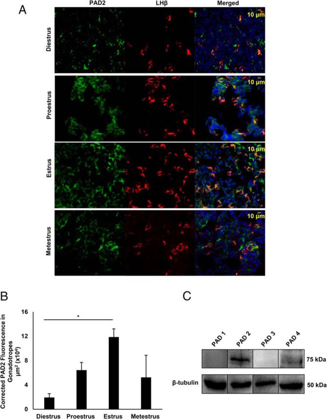
Figure 2. PAD2 expression in mouse gonadotrope cells is highest during estrus.
A, Female mice were estrous cycle staged by vaginal cytology and pituitary glands were collected on the morning of diestrus, proestrus, estrus, and metestrus. Pituitaries were fixed, sectioned frozen (16 μm) and probed with appropriate anti-PAD2 (green) and anti-LHβ (red) antibodies and stained with DAPI (blue) to label nuclei. Tissues were imaged with a Zeiss LSM710 confocal microscope at ×40 resolution. B, Three independent pituitary tissue sections from each stage of the estrous cycle were examined for corrected total fluorescence micrometer square (μm2) of PAD2 in gonadotropes using the region of interest (ROI) feature in ImageJ software. Means were separated using Tukey's HSD, asterisks indicate significant differences (*, P < .05), and error bars are SEM. C. Mice were estrous cycle staged by vaginal cytology and pituitary glands collected during estrus. Protein concentrations of female mouse pituitary lysates were determined and equal concentrations loaded and examined by Western blotting. Membranes were probed with anti-PAD1, anti-PAD2, anti-PAD3, and anti-PAD4 antibodies or with anti-β-tubulin as a loading control.

