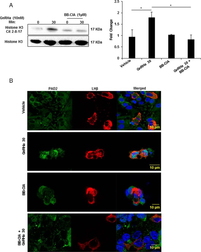
Figure 5. PAD inhibition decreases GnRHa-induced histone citrullination in LβT2 cells.
A, LβT2 cells were pretreated for 12 hours with DMSO or 1μM BB-ClA, then treated with either vehicle or 10nM GnRHa for 0 and 30 minutes. After treatment, histones were purified by acid extraction and quantified, and equal concentrations were examined by Western blotting. Membranes were probed with an antihistone H3 antibody that detects citrullination at arginine residues 2, 8, and 17 and antihistone H3 as a loading control. Bar graphs on the right show quantitative analysis of the Western blottings (n = 3) conducted using Bio-Rad ImageLab software and normalized to total histone H3 levels. Means were separated using Tukey's HSD, asterisks indicate significant differences (P < .05), and error bars are SEM. B, Mice were estrous cycle staged by vaginal cytology and pituitary glands collected during estrus. Pituitaries were explanted, dispersed, and cultured for 12 hours with DMSO or 1μM BB-ClA. Cells were next treated with vehicle or 10nM GnRHa for 30 minutes, then fixed and probed with anti-PAD2 (green) and anti-LHβ (red) antibodies and stained with DAPI (blue). Tissues were imaged with a Zeiss LSM710 confocal microscope using a ×40 objective.

