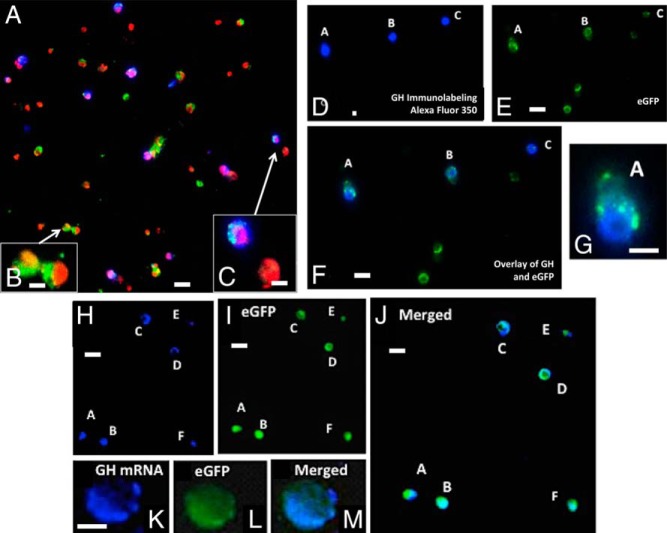
Figure 2.
A, Immunolabeling for GH with Alexa Fluor 350 on a field of mixed pituitary cells from a heterozygous female mouse bearing rGHp-Cre, one floxed allele of Lepr exon 1, and the floxed tdTomato-eGfp Cre-reporter. After immunolabeling, the cells on the coverslip were mounted in media containing propidium iodide (red fluorescence; Vector Laboratories) to detect nuclei. Thus, the following color combinations can be seen: green, eGFP only; blue, GH only; cyan, GH and eGFP; orange/red, all nuclei and tdTomato cytoplasm; purple, GH labeling in cells with red nuclei. B, Cells bearing eGFP without GH labeling fluoresced green with orange/red nuclei. These two cells were not labeled for GH, as expected for mutant somatotrope Lepr-null populations. C, One of these cells shows that GH immunolabeling introduced blue fluorescence, which turned cyan, when overlaid with green. An overlay of blue GH immunolabeling also rendered the nuclei purple. The other cell is a red tdTomato cell that contains no GH. D–F, A field showing five eGFP cells from a mutant mouse illustrating the lack of GH stores in a subset of these cells, as reported previously (8). Three of the cells in this field show immunolabeling for GH (cells A, B, and C in field D or the overlay in field F). G, A higher magnification of cell A from this field. H–J, A field containing six eGFP cells from these mutants, which have been labeled for Gh mRNA with in situ hybridization. As reported previously, most of the somatotropes that are Lepr null have some labeling for Gh mRNA (10). This figure confirms these findings, showing Gh mRNA (blue fluorescence, Alexa-fluor 350; panel H) in all of the eGFP cells (panel I). The overlay of the two fields turns the labeling cyan (panel J). K–M, A higher magnification of a cell labeled for Gh mRNA, eGFP, and the overlay of both labels. Bar, 30 μm (panels A, D, E, F, H, I, and J); 10 μm (panels B, C, G, and K).

