Figure 3.
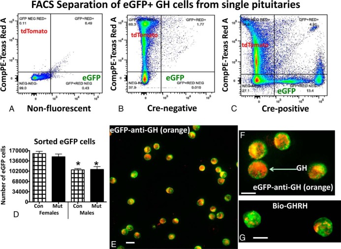
A–C, FACS report of the sorting of cells from nonfluorescent (A), Cre-negative fluorescent (B), and Cre-positive (C) fluorescent reporter mice. FACS is gated to separate tdTomato (532 nm, green laser) and eGFP (488 nm, blue laser) cells. Fluorescence strength is shown along the y- and x-axis for each of the fluorophores. D, Graph of average numbers of eGFP cells collected after FACS and show that females have a higher yield than males. Con, control; Mut, somatotrope Lepr-null mutant. *, Lowest values showing only a sex difference (ANOVA/Tukey's multiple comparisons test; n = individual fractions from 38 control females, 23 mutant females; 28 control males, 16 mutant males). E and F, eGFP fraction immunolabeled for GH with 1:30 000 anti-GH detected by biotinylated goat antirabbit IgG and streptavidin Dylight 549 (red-orange fluorescence). GH labeling is detected in most eGFP cells. G, eGFP cells were treated with biotinylated GHRH for 10 minutes and then fixed and labeled with streptavidin Dylight 549. Most eGFP cells show binding in peripheral patches at the periphery. Bar, 12 μm.

