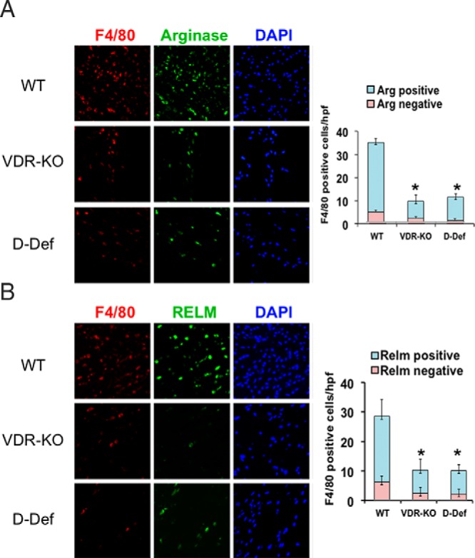
Figure 1.

Ligand-dependent actions of the VDR regulate M2 macrophage response to cutaneous injury. IHC analyses for F4/80 were performed on 2-day wounds of WT, VDR KO, and vitamin D-deficient (D-Def) mice to evaluate M2 macrophages. Markers of M2 polarization, arginase (A) and RELMα (B), were also evaluated. Graphs represent the number of F4/80+ cells per high-power field that were found to be arginase (Arg) and RELM positive or negative. Data represent those obtained from five wounds per condition. *, P < .0001 vs WT by ANOVA (VDR KO vs D-Def, P = NS). DAPI, 4′,6′-diamino-2-phenylindole.
