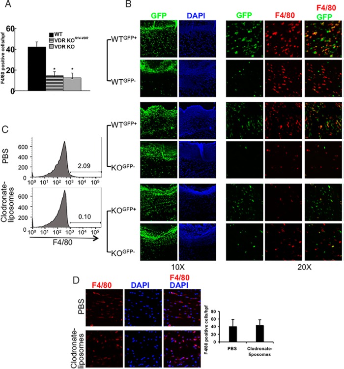
Figure 2.
Cutaneous wound granulation tissue macrophages are of local tissue origin. A, Restoring VDR expression in keratinocytes of VDR KO (VDR KOK14VDR) mice does not normalize cutaneous wound macrophage number. Data represent the mean and SD of the number of F4/80-positive macrophages per high-power field in five wounds per genotype. *, P < .0001 vs WT by ANOVA (VDR KO vs VDR KOK14-VDR, P = NS). B, Parabiosis studies were performed in WT and VDR KO mice differing in GFP status. GFP fluorescence and DAPI nuclear staining of the clot and granulation tissue of day 2 wounds are shown under low magnification (×10). GFP and F4/80 dual immunofluorescence is shown under high power (×20). Data are representative of those obtained with five parabiotic pairs per condition. C, Flow cytometric analyses demonstrate depletion of circulating F4/80-positive cells by clodronate liposomes. D, F4/80 IHC assays of wound macrophages in clodronate liposome- and PBS-injected mice. Graph represents the mean and SD of the number of F4/80-positive cells/hpf. Data are representative of those obtained in six wounds per genotype. DAPI, 4′,6′-diamino-2-phenylindole.

