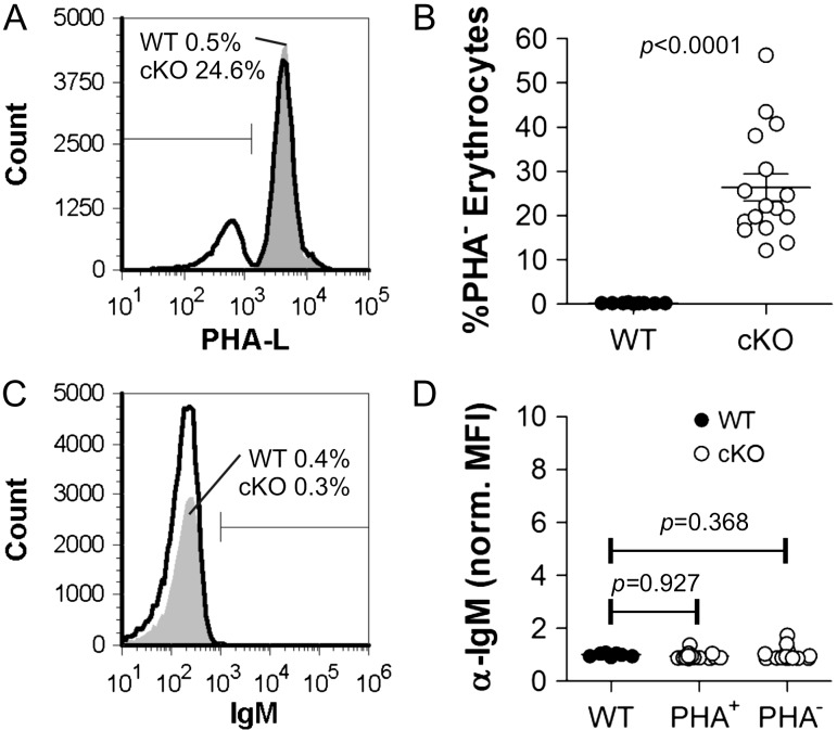
Fig. 3.
DC-cKO erythrocytes (cKO) show Mgat2 ablation but not autoantibody deposition. Erythrocytes were collected from WT (CD11c-CRE-GFP) or cKO mice and stained with PHA-L and anti-mouse IgM. (A, B) Approximately 30% of erythrocytes from cKO mice show a lack of complex N-glycans (n = 16; P < 0.0001). (C, D) None of the erythrocytes for either WT or cKO mice showed detectable auto-IgM antibody deposition, in contrast to Mgat2−/− erythrocytes from M-cKO mice previously reported (Ryan et al. 2013) (n = 16; P = 0.368, 0.927).

