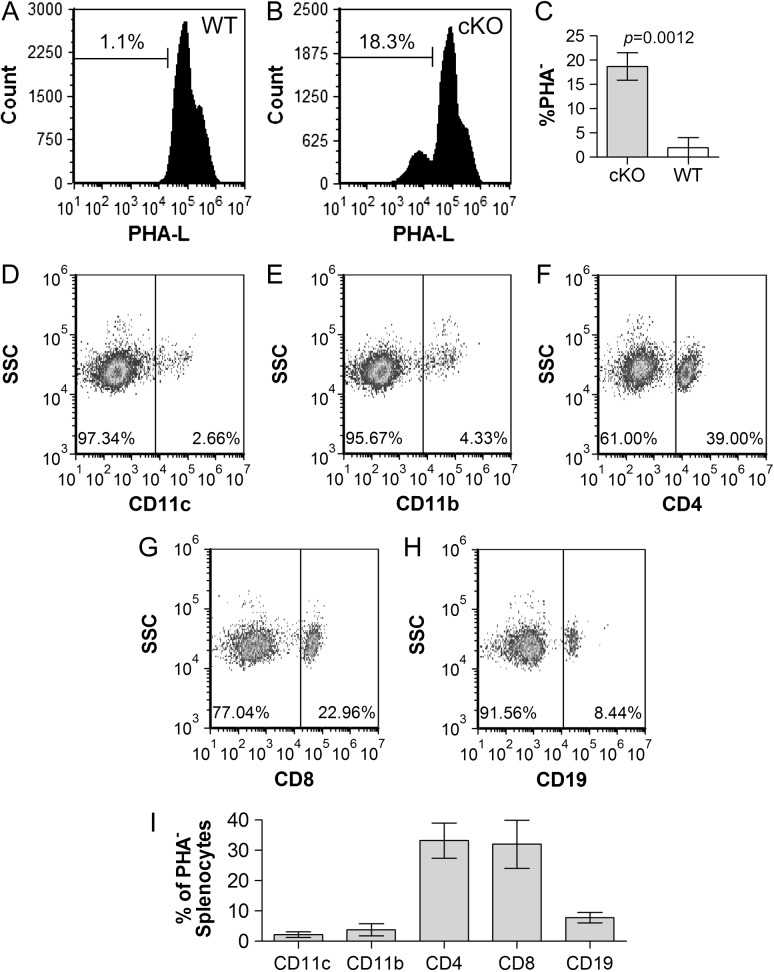
Fig. 7.
T cells are the most abundant splenic Mgat2-null cells. WT (A) and DC-cKO (B) Spls were harvested and single cells stained with PHA-L. PHA-L− cells from DC-cKO mice (C) were also stained with antibodies against CD11c (D), CD11b (E), CD4 (F), CD8 (G) or CD19 (H) and plotted against side-scatter. Data from replicates are also shown (I). In the Spl, over 60% of all Mgat2-ablated cells were T cells (CD4+ and CD8+), with CD11c+ cells accounting for only 2.6% of the total PHA-L− cells (n = 3).

