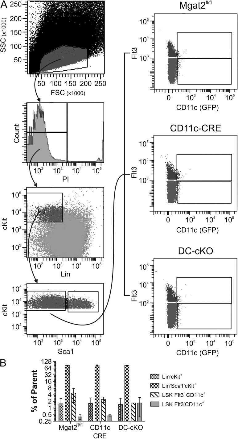
Fig. 8.
HSCs show no defects or CD11c expression. Bone marrow from both parental mouse strains (CD11c-CRE-GFP and Mgat2fl/fl) and DC-cKO mice was harvested and stained with propidium iodide (PI) and antibodies against lineage markers (Lin), cKit, Sca1 and Flt3. GFP fluorescence was used as a marker of CD11c expression. (A) The gating scheme for analysis is shown, which revealed a lack of GFP signal in the HSC compartment. (B) Quantitation of each HSC compartment is shown, indicating a lack of HSC compartment changes in cell proportion associated with loss of Mgat2 (n = 3; P > 0.05 between genotypes for each cell lineage).

