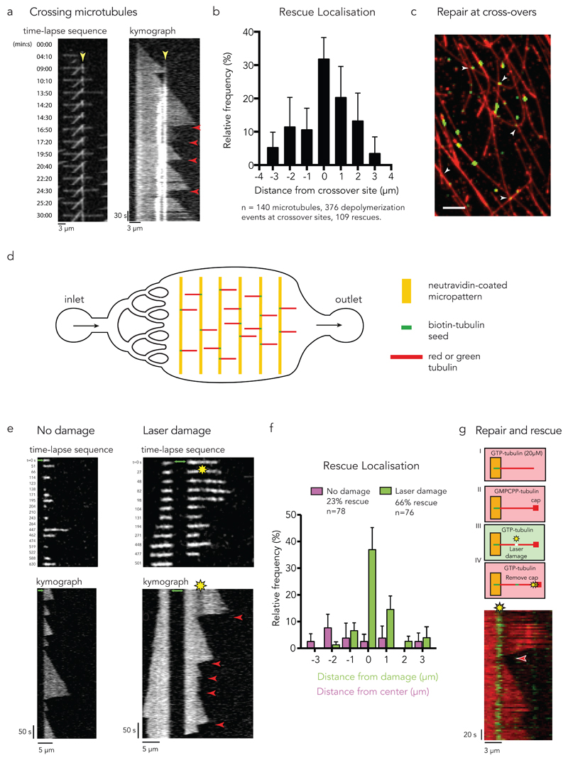
Figure 4. Microtubule self-repair induces rescue events in vitro.
(a) Rescue at crossing microtubules. Time-lapse sequence of 3 microtubules crossing each other. The kymograph highlights the crossing sites (yellow arrow-head pointing at the bright white vertical lines) and the occurrence of multiple rescue events at this site (red arrow-heads).
(b) The graph shows the frequency of rescue events for crossing microtubules as a function of distance from the crossing site. Data represent mean +/- s.d from n=8 independent experiments.
(c) Repair at crossing microtubules. Observation of the incorporation of green tubulin dimers along red microtubules. White arrow-heads point at crossing sites where accumulation of green tubulin was detected. Image is representative of 3 independent experiments. Scale bar 5 µm.
(d) Illustration of the microfluidic device. Short biotinylated microtubule seeds were fixed on neutravidin coated micropatterns and elongated using red or green free tubulin. To exchange or remove the solution of free tubulin, a flow was induced parallel to the microtubules.
(e) Photo damage sites can induce rescue. The image sequences and kymographs show microtubule dynamics with (right) and without (left) laser-induced damage. The green arrows indicate the seed. Red arrow-heads indicate rescue events.
(f) The graph shows the frequency of rescue events for photo damaged microtubules as a function of distance from the center of the damage (green bars) and for microtubules without damage as a distance from the center of the observed microtubule (magenta bars). Data represent mean values +/- s.d from n= 4 independent experiments.
(g) Tubulin incorporation at photodamaged sites is associated with rescue. Green microtubule seeds were elongated with red free tubulin (step I). A GMPCPP cap was grown at the microtubule tip to avoid spontaneous depolymerization (step II). Photo damage was induced in the presence of green tubulin (step III). Depolymerization was initiated by removing the GMPCPP cap with a laser pulse at high intensity (step IV). The kymograph shows rescue (red arrow-head) at the damaged site where green tubulin was incorporated.
Image is representative of 4 independent experiments.

