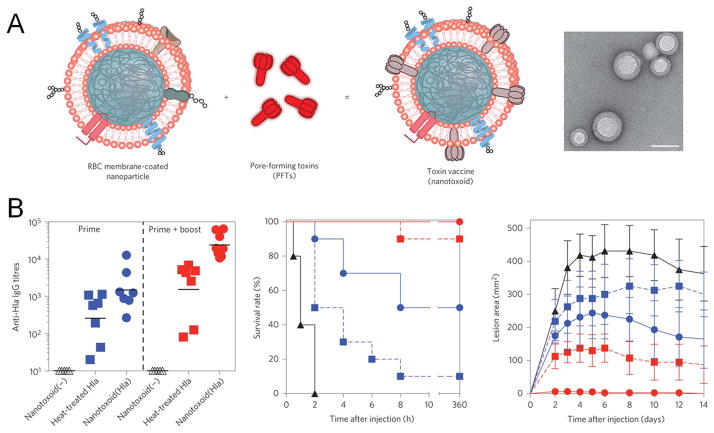
Figure 5.
Erythrocyte membrane-coated PLGA nanoparticles for vaccine delivery of staphylococcal α-haemolysin (Hla). (A) Schematic illustration and an image by transmission electron microscope of the nanotoxoid. Scale bar, 80 nm. (B) The nanotoxoid vaccine enhanced humoral immune responses and protected mice against toxin challenge. Empty triangles, vaccine particles without the antigen; solid triangles, unvaccinated control; blue squares, single dose of the heat-inactivated Hla; blue spheres, single dose of the nanotoxoid; red squares, three doses of the heat-inactivated Hla; red spheres, three doses of the nanotoxoid. Reproduced with permission from ref. 112.

