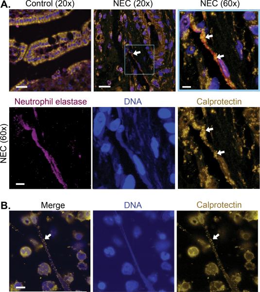
Figure 4. NETs contribute to intestinal injury associated with NEC and are decorated with calprotectin protein.
(A) We used immunohistochemical techniques to compare calprotectin protein expression in gastrointestinal tissue samples collected from 5 different infants who underwent surgical treatment for NEC and 6 control infants operated on for indications other than NEC. Representative images of each are shown. Calprotectin is seen in gold fluorescence. Neutrophil elastase, a protein known to be expressed on NETs, is seen in magenta fluorescence. Nuclear and NET-associated DNA is shown in blue fluorescence. A NET is seen in the intestinal tissue of an infant with NEC (arrows), while no NETs or neutrophil elastase staining were seen in the control tissues. The magnified image of the highlighted box, complete with the three separate fluorescent channel images, demonstrate neutrophil elastase, DNA, and calprotectin staining consistent with NET formation. The images highlighting DNA and neutrophil elastase have been brought up to brighter exposures for the purpose of clearer demonstration. (B) NET formation was detected using immunocytochemistry with the same primary antibodies used to detect calprotectin and DNA employed in panel A. Freshly isolated adult PMNs were treated with LPS (100 ng/mL) for 1 hour to stimulate NET formation. Again, gold fluorescence denotes calprotectin protein expression and blue fluorescence denotes DNA. These images are representative of 6 different replicates using PMNs isolated from 6 different healthy adults. Bar, 20μm on A row 1, images 1 and 2, 5μm on A row 1, image 3 and row 2, and 10μm on B.

