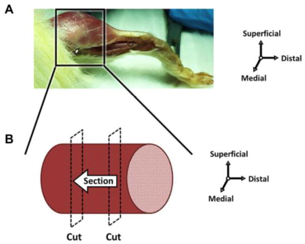
Figure 1. Schematic of muscle harvest for immunohistochemistry.
(A) Muscle tissue was excised posterior to the tibia and proximal to the myotendinous junction, and then flash frozen. (B) Prior to sectioning, distal and proximal portions of the muscle tissue were removed, effectively isolating the muscle belly. The muscle was sectioned perpendicular to the long axis of the fibers from the distal to proximal direction.

