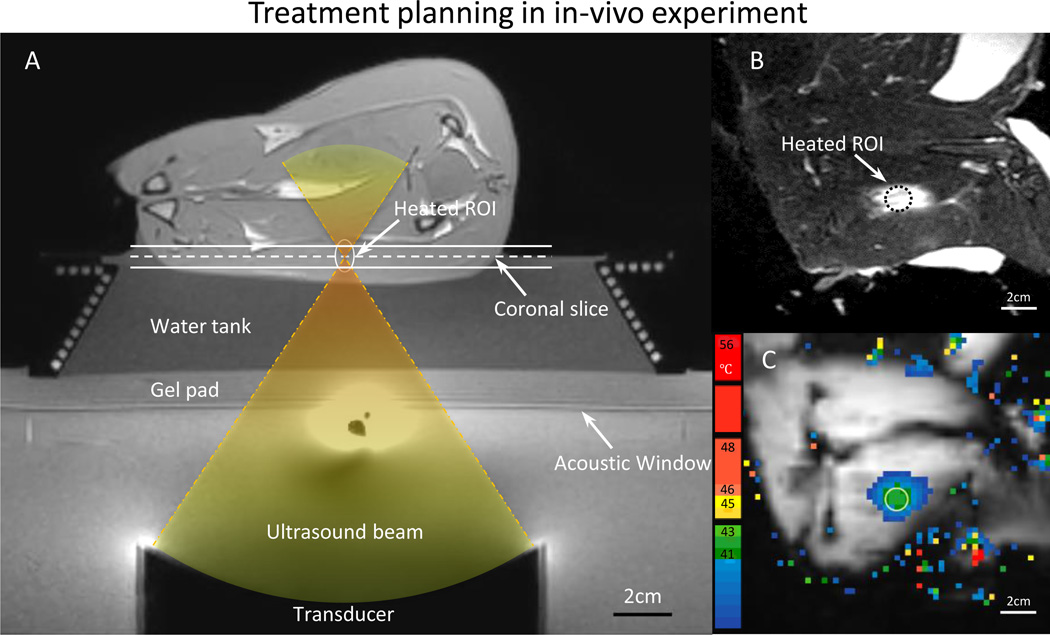
Fig. 3.
A) T1-weighted image along the ultrasound beam axis shows the experimental setup for performing hyperthermia in a rabbit model. A gel pad and a water tank were placed on top of the acoustic window of the clinical MR-HIFU system to elevate the animal to the location of the ultrasound focus. The conical water tank also maintained the body temperature of the animals. B) T2-weighted image transverse to the beam axis (along the dashed line in A) shows the VX2 tumor and the location of heating. C) The spatial temperature distribution measured midway during treatment shows localized heating within the target area, well maintained in the desired range of 41–43 °C.

