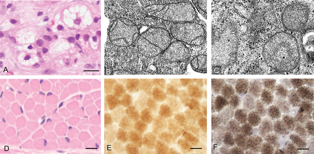
Figure 1.
Histological and ultrastructural evidence for hepatic microsteatosis and mitochondrial structural defects. (A) Hematoxylin and eosin-stained sections of liver biopsy show microsteatosis (multiple vacuoles) in the cytoplasm (scale bar 10µm). (B–C) Electron microscopic examination of liver shows structural abnormalities in mitochondria (B) 20,000× and (C) 30,000×. (D) Muscle biopsy of right quadriceps muscle stained with hematoxylin and eosin (scale bar 20µm). (E–F) Staining for (E) COX and (F) combined SDH and COX, these stains showed no evidence of COX deficient fibers or ragged-blue fibers (scale bar 10µm).

