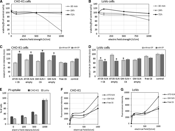
Fig. 5.
Top row the effect of EP parameters on cell viability (MTT test): a CHO-K1 cells; b LoVo cells. Middle row the viability of cells after treatment with free C6, empty, and C6-loaded nanoparticles (GM-SLN and ATO5-SLN); SLNs and free C6 added before EP: c CHO-K1 cells, d LoVo cells. Bottom row FACS analysis, uptake of PI and free C6, added before EP, and SLNs added post-EP: e CHO-K1 and LoVo cells—dependency of PI uptake (percent of cells) on EP parameters; f CHO-K1 cells—uptake of free and encapsulated C6 (fluorescence intensity); g LoVo cells—uptake of free and encapsulated C6 (fluorescence intensity)

