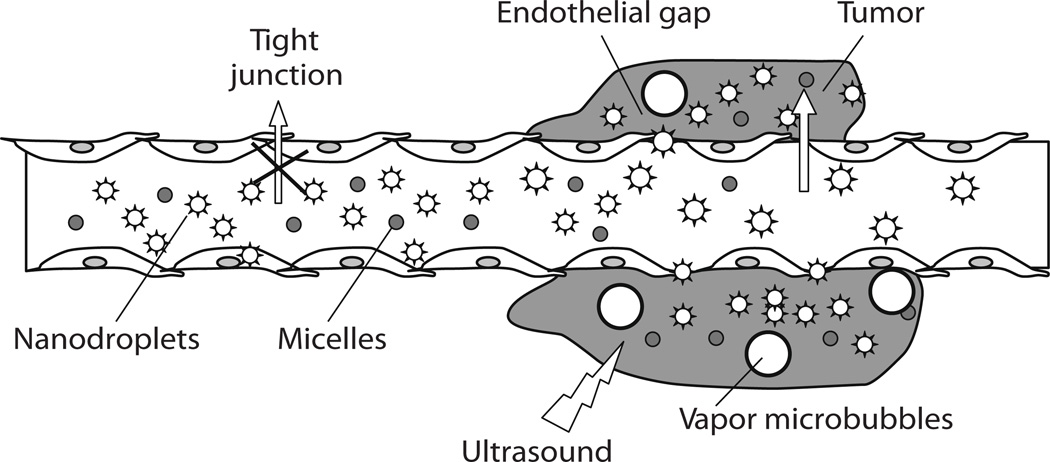
Fig. (5).
Schematic representation of passive drug targeting through the defective tumor microvasculature using an echogenic drug delivery system. The system comprises micelles (small circles), PFC nanodroplets (stars), and PFC microbubbles (large circles). Lipophilic drugs can be localized in the micelle cores and in the walls of nanodroplets/microbubbles. Tumors are characterized by defective vasculature with large gaps between the endothelial cells, which allows extravasation of drug-loaded micelles and small nanodroplets into the tumor interstitium. Primary microbubbles are formed from the vaporization of nanodroplets due to hyperthermia or ultrasound, and larger microbubbles appear due to coalescence of the primary microbubbles. Rapoport N, Gao Z, Kennedy A, “Multifunctional nanoparticles for combining ultrasonic tumor imaging and targeted chemotherapy”, J Natl Cancer Inst, 2007, Vol. 99, 1095–106, by permission of Oxford University Press.

