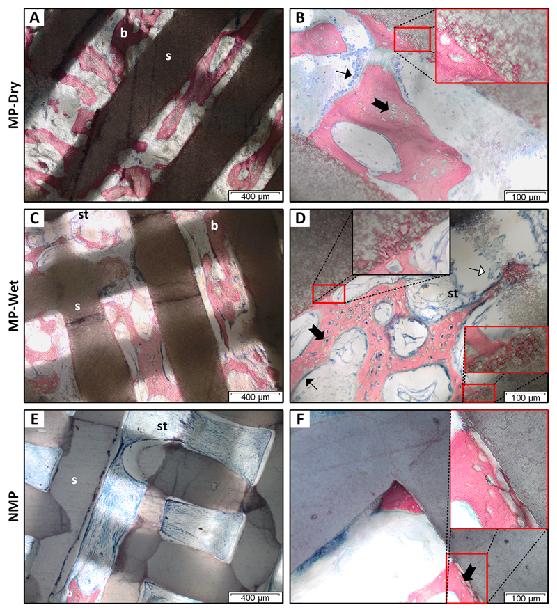
Figure 2. Tissue and cells in the scaffold macro- and micropores.
Histology slices were taken at the center of the scaffolds. Samples were stained with Stevenel’s blue and counterstained with picro-fuchsin. Bone was pink/red; soft tissue, osteoid, cell cytoplasm were light blue; and cell nuclei were dark blue. Mineralized bone (b) was observed in macropores between scaffold rods (s) for all three groups. Fibrous soft tissue (st) was prevalent in the center of NMP scaffolds (E). Osteoblast-like cells (➙) lined mineralized bone in the macropores (B, D). Osteocytes (➞) were in lacunae (B, D, F). Osteoclast-like cells (⇾) were found on rod and bone surfaces in some areas (D). In the macropores of MP scaffolds, mineralized bone is anchored in rods (B, D, insets). In NMP, bone is not anchored (F, inset). Soft tissue in MP-Wet and NMP is contracted and frequently not in contact with the rods (C- E).

