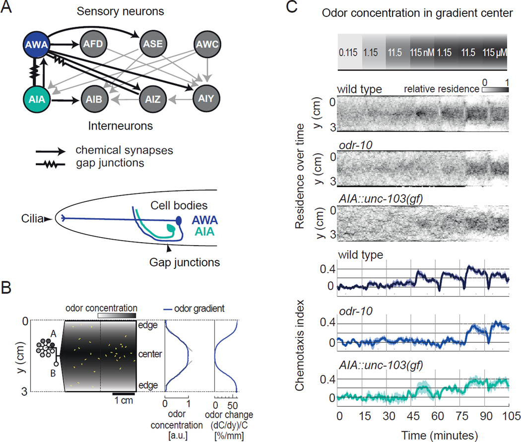
Figure 1. A neural circuit for chemotaxis towards defined concentrations of diacetyl.
a) The AWA circuit. Top, part of the C. elegans wiring diagram (White et al., 1986), emphasizing synaptic connections between AWA olfactory neurons and other sensory neurons and interneurons. Each sensory neuron has additional targets, and each interneuron integrates input from additional sensory neurons. Bottom, schematic illustration of AWA and AIA. AWA detects odors via cilia on the distal sensory dendrite and forms axonal gap junctions with AIA interneurons.
b) Schematic of microfluidic behavioral device in which sigmoidal odor gradients of known diacetyl concentrations are delivered to freely moving animals. Diacetyl concentrations at the outer edges are held constant at 0.115 nM via inlet B. Peak odor concentration is selected using a computer controlled distribution valve via inlet A. Middle, odor concentration profile. Gradient in arbitrary units (a.u.) scales with peak odor concentration, and the slope of much of the gradient is near-linear. Right, concentration change per mm experienced by an animal moving toward the center of the arena; animals move at about 0.2 mm/second. See also Supplemental Movie 1.
c) Behavior of wild-type and mutant animals in diacetyl gradients. The peak diacetyl concentration was increased ten-fold as indicated every 15 minutes (top). Middle three panels, distribution of animals in the device expressed as a density function of residence over time. Bottom three panels, a mean chemotaxis index represented the location of animals in the device during 2 s time bins (continuous scale from −1 at device edge to +1 at device center). odr-10(ky32) mutants lack the AWA diacetyl receptor. unc-103(gf) strains express a hyperactive K+ channel in the AIA neurons under the gcy-28d promoter. Shading represents s.e.m. n = 4–6 assays with 20–30 animals per genotype.

